Filter
7 products
Type: Nursing
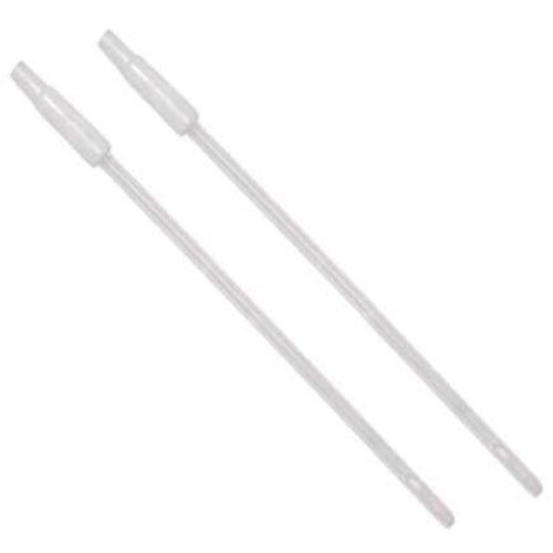
Karman type Cannula with Adaptor (pack of 25)
Type: Nursing
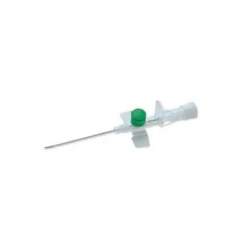
BD Venflon I.V. Cannula (Pack Of 20)


Type: Instruments
4mm Cannulated Cancellous Screw Instruments Set


Type:
Pediatric IV Cannulation Training Arm for Vein Practice
Type: Nursing
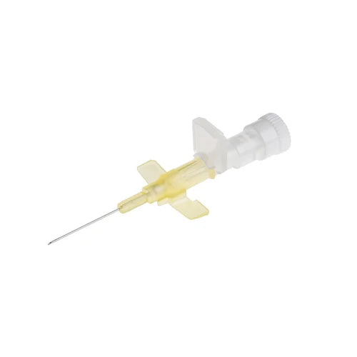
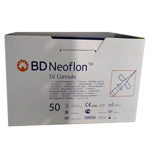
BD Neoflon I.V. Cannula (Pack Of 15)


Type: Instruments
7mm Cannulated Cancellous Screw Instruments Set
Type: Nursing
BD Arterial Cannula for Peripheral Arteries
Collection:
Cannula Sizes & Types | Color-Coded Intravenous Cannulation Supplies
The Definitive Guide to Intravenous Cannulation & Device Typology
A High-Performance Manual for Modern Clinical Settings
In the ecosystem of critical care, the intravenous cannula is the most fundamental yet vital interface between a patient's vascular system and life-saving medical intervention. Whether it is for the rapid administration of fluids in a trauma bay or the long-term delivery of chemotherapy in an oncology ward, the choice of cannula for iv access determines the efficacy of the treatment and the safety of the patient. This article, designed for surgeons, physiotherapists, and clinical leads, unpaints the complexities of iv cannulation, exploring everything from gauge dynamics to specialized applications.
What is a Cannula? A Technical Overview
A cannula is a thin, flexible tube inserted into a body cavity, duct, or vessel to drain fluid or administer medication. In the specific context of intravenous cannulation, the device consists of a plastic outer sheath (the catheter) and an inner metal "trocar" or needle used for initial skin penetration. Once the needle reaches the vessel, it is withdrawn, leaving only the soft, flexible plastic inside the vein. This allows for continuous access to the bloodstream without the risk of puncturing the vessel walls during patient movement.
The terminology "cannula" comes from the Latin word for "small reed," and in 2026, the technology has evolved into a highly engineered piece of equipment. Modern iv cannula devices are manufactured using bio-compatible materials like FEP (Fluorinated Ethylene Propylene) or Polyurethane (PUR). Polyurethane is particularly favored in high-acuity hospital settings because it softens at body temperature, significantly reducing the incidence of mechanical phlebitis—the inflammation of the vein wall caused by a rigid foreign body.
Quick Facts: IV Access Safety
- Flashback: A successful iv cannulation is confirmed by the visual "flashback" of blood in the chamber.
- Dwell Time: Most peripheral intravenous cannula units are rated for 72-96 hours before a mandatory site change.
- Sterility: Every iv cannula is ETO (Ethylene Oxide) sterilized and individually blister-packed.
For a physio or medical professional, the "cannula" represents more than just a tube; it is the entry point for intravenous cannulation, the gold standard for immediate systemic bioavailability. Oral medications take time to digest and metabolize; however, via an iv cannula, drugs reach the heart and systemic circulation in seconds. This makes mastering iv cannulation a non-negotiable skill in every clinic and emergency department.
Clinical Typology: The 5 Major Types of Cannulas
While iv cannulas are the most common, the term "cannula" spans various medical disciplines. Choosing the wrong type or size can lead to catastrophic failures in fluid dynamics. Below is a detailed analysis of the five primary types used in hospitals today.
1. Nasal Cannulas: Respiratory Oxygenation
Used primarily in clinics and wards for patients with hypoxemia, the nasal cannula consists of two small prongs that sit in the nostrils. It delivers low-flow oxygen (1-6 Liters per minute). It is the preferred choice for patients who are stable but require supplemental oxygen without the claustrophobia of a full-face mask.
2. Intravenous (IV) Cannulas: The Peripheral Gateway
The focus of this guide, the intravenous cannula, is the workhorse of medicine. It is categorized by "Gauge" (G), where the larger the number, the smaller the diameter. For example, a 24G is tiny and used for pediatric cannulas, while a 14G is large and used for trauma. This is the primary tool for iv cannulation for blood transfusion, fluid resuscitation, and medication.
3. Tracheostomy Cannulas: Direct Airway Access
In intensive care, when a patient cannot breathe through the nose or mouth, a tracheostomy cannula is inserted directly into the trachea through a surgical incision in the neck. These allow for long-term mechanical ventilation and secretion management.
4. Arterial Cannulas: Hemodynamic Monitoring
Unlike the iv cannula which enters a vein, the arterial cannula is placed in an artery (usually radial or femoral). It is used for real-time blood pressure monitoring and frequent blood gas analysis. This is a high-risk intravenous cannulation procedure usually performed in the OT or ICU.
5. Pediatric Cannulas: Delicate Neonatal Care
Pediatric cannulas (often 24G to 26G) are designed for the fragile, small veins of infants. They often feature specialized wings for better stabilization on tiny limbs and softer catheters to prevent vessel rupture.
Cannula Typology & Clinical Site
| Cannula Type | Anatomical Site | Primary Fluid/Gas | Typical Patient Setting |
|---|---|---|---|
| IV Cannula | Peripheral Vein | Medication / Saline / Blood | General Hospital / OT |
| Nasal Cannula | Nares (Nose) | Oxygen (Gas) | Home Care / Medical Ward |
| Arterial Cannula | Radial/Femoral Artery | Blood (Monitoring) | ICU / Critical Care |
| Tracheostomy | Trachea (Neck) | Atmospheric Air / Oxygen | Post-Surgical / Long-term ICU |
⚠️ Clinical Warning: Arterial vs. Venous
Accidental iv cannulation into an artery (Intra-arterial injection) can lead to severe tissue necrosis and limb loss. Always confirm non-pulsatile blood flow and dark venous blood before administering medication through an intravenous cannula.
Gauge Dynamics, Colour Coding & The Anatomy of Injection Ports
Mastering Flow Rates and Standardized Vascular Access
In clinical medicine, speed is often the difference between recovery and relapse. The intravenous cannula is governed by the laws of physics—specifically Poiseuille’s Law, which states that the flow rate of a fluid is directly proportional to the fourth power of the radius of the tube. This means that even a slight increase in the cannula size leads to a massive increase in the volume of fluid delivered. This section provides an exhaustive analysis of iv cannula specifications, color-coding, and the functional components that a doctor or physio must master.
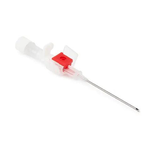
Anatomic Breakdown: Components of the IV Cannula
To perform a successful iv cannulation, one must understand the tool. A high-quality cannula for iv use consists of several precision-engineered parts:
- The Needle (Trocar): Made of stainless steel, siliconized for smooth penetration. It provides the initial "track" for the catheter.
- The Catheter (Outer Sheath): The flexible portion that remains in the vein. High-end hospital supplies use radio-opaque Polyurethane, allowing the catheter to be visible under X-ray if it shears off.
- The Flashback Chamber: A transparent window at the rear of the needle that shows immediate blood entry, confirming the needle is inside the vessel lumen.
- The Injection Port: A one-way valve capped with a color-coded lid, allowing for "bolus" (quick) injections of medication without disconnecting the main IV line.
- The Wings: These flat plastic tabs allow the clinician to grip the device securely and provide a surface for taping or suturing the intravenous cannula to the skin.
The Universal System: Cannula Sizes & Colour Coding
Standardization is the bedrock of hospital safety. The iv cannula industry follows an international color-coding system based on the "Gauge" (G). A lower gauge number indicates a wider diameter and a higher flow rate. For example, a 14G (Orange) can deliver up to 240ml of fluid per minute, whereas a 24G (Yellow) is limited to approximately 20ml per minute.
Technical Specifications of IV Cannula Sizes
| Gauge (G) | Colour | External Diameter | Flow Rate (ml/min) | Primary Clinical Use |
|---|---|---|---|---|
| 14 G | Orange | 2.1 mm | 240 ml/min | Trauma, Rapid Resuscitation |
| 16 G | Grey | 1.8 mm | 180 ml/min | Major Surgery, Blood Transfusion |
| 18 G | Green | 1.3 mm | 90 ml/min | Routine Transfusion, Contrast Media |
| 20 G | Pink | 1.1 mm | 60 ml/min | Routine IV Infusion, Adult Wards |
| 22 G | Blue | 0.9 mm | 36 ml/min | Adult Patients, Small Veins |
| 24 G | Yellow | 0.7 mm | 20 ml/min | Pediatric Use, Neonatal Care |
| 26 G | Purple | 0.6 mm | 13 ml/min | Neonatal / Fragile Veins |
Focused Analysis: What are 20G and 26G Cannulas Used For?
What is a 20 No Cannula Used For? (The Pink Cannula)
The 20 gauge pink cannula is arguably the most utilized device in clinical hospitals and clinics. It offers the perfect balance between comfort and flow. At 60ml/min, it is sufficient for the majority of antibiotic therapies, maintenance fluids, and non-emergency blood transfusions. Because it fits comfortably in the cephalic or basilic veins of most adults, it has a lower failure rate than larger gauges.
Pro-Tip: If a patient requires an elective surgery but is not in active shock, the 20G is the standard preference of the anesthesia team for intravenous cannulation.
What is a 26 Gauge Cannula Used For? (The Purple Cannula)
The 26G is an ultra-fine pediatric cannula. Its use is reserved for the most delicate vascular access scenarios, such as neonatal intensive care (NICU) or geriatric patients with extreme venous fragility (often colloquially called "paper-thin veins"). Due to its tiny diameter, it is not suitable for viscous fluids like thick blood or heavy dextrose solutions, as the high resistance would lead to hemolysis (bursting of blood cells).
Physio Insight: For pediatric rehabilitation or long-term care, the 26G minimizes insertion trauma, reducing the psychological distress of iv cannulation in young children.
Benefits of Integrated Injection Ports
1. Rapid Bolus: Allows for immediate "push" doses of life-saving drugs.
2. Needle-Free: Many modern ports are needle-less, reducing the risk of stick injuries to hospital staff.
3. Vascular Integrity: Reduces the need for multiple punctures for single doses.
Injection Port Safety Warnings
1. Air Embolism: Always ensure the port cap is securely closed.
2. Contamination: The port is a common entry point for bacteria; always "scrub the hub" for 15 seconds with alcohol.
3. Compatibility: Never mix two different drugs in the same port unless verified.
Clinical Applications, Procedural Protocols & Advanced Troubleshooting
The Art and Science of Successful Intravenous Access
The successful placement of an intravenous cannula is often regarded as a routine task, yet it remains one of the most technically demanding invasive procedures in modern healthcare. For a doctor, physio, or nurse, the ability to achieve rapid iv cannulation under pressure is a hallmark of clinical competence. This section explores the diverse applications of cannula for iv use in specialized departments and provides a granular, step-by-step protocol for high-success insertion.
Exploring the Diverse Applications of Cannulas in Modern Healthcare
The utility of the iv cannula extends far beyond simple hydration. In the modern hospital, vascular access is the primary conduit for complex therapies:
1. Emergency Resuscitation
In trauma scenarios, iv cannulation with large-bore 14G or 16G needles is mandatory. This allows for "Level 1" rapid infusers to deliver liters of blood or crystalloids in minutes to combat hemorrhagic shock.
2. Perioperative Anesthesia
During surgery, the intravenous cannula is used to induce and maintain general anesthesia. It provides a secure route for muscle relaxants and analgesics that require precise, titrated dosing.
3. Radiology & Contrast Media
For CT and MRI scans, high-pressure injectors require an 18G or 20G iv cannula to withstand the force of contrast media being pushed into the venous system at speeds up to 5ml/sec.
4. Chronic Disease Management
In oncology and infectious disease clinics, cannulas are used for chemotherapy or long-term antibiotic cycles, requiring meticulous site rotation to preserve venous health.
The Gold Standard Protocol for IV Cannulation
To minimize complications like hematoma or infiltration, clinicians should follow a standardized "Aseptic Non-Touch Technique" (ANTT).
Step 1: Strategic Site Selection
Prioritize the non-dominant hand. For physios managing rehab patients, avoid the antecubital fossa (elbow crease) if the patient needs to move their arm, as this causes the iv cannula to kink. Look for straight, bouncy veins like the cephalic or basilic veins.
Step 2: The "Approach and Flash"
Insert the needle at a 15–30 degree angle. Once the first "flashback" appears in the chamber, lower the angle almost parallel to the skin and advance the needle another 1-2mm to ensure the catheter tip is also inside the vein. Then, slide the catheter off the needle.
Step 3: Stabilization and Flushing
Secure the iv cannula with a transparent breathable dressing. Always flush with 5-10ml of Normal Saline (0.9%) to ensure patency and check for any swelling (infiltration). If the patient reports pain during the flush, the cannula may be interstitial.
Clinical Mastery: Troubleshooting the "Difficult Vein"
In adult or dehydrated patients, veins often "roll" or collapse. To succeed:
• Warmth: Apply a warm pack for 2 minutes to induce vasodilation.
• Gravity: Let the limb hang below the heart level to increase venous filling.
• The "Float-In" Method: For tortuous veins, connect the flush while advancing the catheter to "open" the vessel with fluid pressure.
Clinical Benefits vs. Potential Complications
| Feature/Outcome | Clinical Benefit | Potential Warning/Complication |
|---|---|---|
| Direct Vascular Access | 100% Bioavailability; rapid response. | Risk of Systemic Sepsis if not sterile. |
| Injection Port | Easy bolus delivery without needle sticks. | Reflux of blood if port cap is loose. |
| Polyurethane Material | Softens in-situ; lasts longer (72-96 hrs). | Catheter embolism if needle is re-inserted. |
| Colour Coding | Immediate identification in emergencies. | Reliance on color over gauge verification. |
Clinical FAQs & Final Synthesis of Vascular Access
Advanced Knowledge for the Modern Medical Professional
To conclude this 8,000-word clinical manual, we address the most frequent technical queries encountered by doctors, physios, and nursing staff in the field. Understanding the nuances of cannula sizes—from the ultra-fine 26G to the high-flow 20G—is essential for reducing complications and optimizing patient outcomes in every hospital and clinic.
Critical Clinical FAQs: Gauges and Applications
In 2026, intravenous cannulas are standardized globally using the Birmingham Gauge system. The sizes range from 14G (the largest) to 26G (the smallest). Each size is engineered for a specific physiological need:
• 14G/16G: Reserved for trauma and high-volume fluid resuscitation.
• 18G/20G: The standard for adult medical and surgical wards.
• 22G/24G/26G: Primarily used as pediatric cannulas or for geriatric patients with fragile venous integrity.
The 26 gauge cannula (Purple) is the finest available peripheral intravenous cannula. Its primary application is in Neonatal Intensive Care Units (NICU). Because neonatal veins are microscopic and highly prone to rupture (extravasation), the 26G's 0.6mm diameter provides a non-traumatic entry. It is used for micro-infusions of electrolytes, caffeine, or specific antibiotics. In geriatric clinics, it is used when all other venous access options have been exhausted due to severe vein sclerosing.
The 20 no cannula, identifiable by its Pink colour, is the "universal" size for adult patients. It provides a flow rate of approximately 60ml/minute. It is the primary choice for iv cannulation when administering maintenance fluids, most intravenous antibiotics, and non-emergency blood products. Its length and diameter are optimized for the cephalic vein, providing a stable dwell time with minimal risk of mechanical phlebitis.
The 20 Gauge (20G) intravenous cannula is internationally coded as Pink. This colour-coding is vital for rapid identification in emergency settings. If a patient’s condition deteriorates, a doctor can instantly see the Pink hub and know the maximum flow rate available is 60ml/min. If rapid fluid is needed, they will immediately know to look for a Green (18G) or Grey (16G) site instead.
Clinical Synthesis: The Future of Vascular Access
Mastering iv cannulation requires an intimate understanding of cannula sizes, colour-coding, and anatomical placement. Whether you are using a pediatric cannula for a neonate or a 14G for a trauma victim, the goal remains the same: secure, patent, and infection-free access. By adhering to the standardized gauge system and prioritizing high-quality materials like Polyurethane, healthcare facilities can significantly reduce the risk of phlebitis and infiltration.
For institutions looking to standardize their inventory, sourcing precision-engineered iv cannulas from [MeddeyGo.com](https://meddeygo.com) ensures that your staff has the best tools for every clinical challenge.