Filter
5 products
Type: Devices
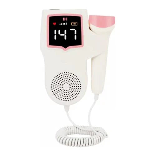
Portable Fetal Doppler 2.0 MHz

Type: Devices
Vcomin Fetal Doppler 300D

Type: Devices
Vcomin Fetal Doppler 200D


Type: MeddeyGo
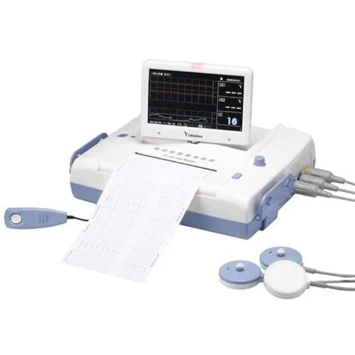
Fetal Monitor with CTG Flight 800
Type: Devices
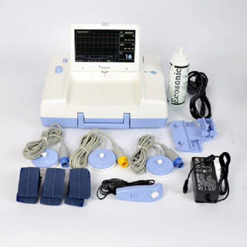
Bistos BT350 Fetal Monitor CTG Machine with 7 inch Screen LED Light Source
Collection:
Fetal Dopplers & Heart Rate Monitoring: The Definitive Clinical Guide
A fetal doppler is an essential handheld ultrasound tool used to detect and monitor the fetal heartbeat during pregnancy. For clinicians and expecting parents alike, this doppler instrument provides the first audible confirmation of a healthy pregnancy. At Meddeygo.com, we prioritize medical-grade precision to ensure that every heartbeat is captured with absolute clarity and safety.
What is a Fetal Doppler Used For?
The primary use of a fetal doppler is to monitor the Fetal Heart Rate (FHR). It works on the "Doppler Effect" principle, sending high-frequency sound waves into the abdomen which bounce off the fetal heart. The shift in frequency is then converted into an audible sound and a digital BPM (Beats Per Minute) reading.
Fetal Heart Rate Monitoring: What Does It Tell?
Monitoring the FHR provides critical insights into the baby's well-being, specifically:
- Oxygenation: A steady heart rate indicates the placenta is providing sufficient oxygen.
- Stress Levels: Significant drops (decelerations) or spikes (tachycardia) can signal fetal distress.
- Reactivity: Normal variations in the heart rate show a healthy developing nervous system.
Is it Safe to Use a Fetal Doppler at Home?
This is a common debate in 2026. While a fetal doppler uses non-ionizing ultrasound (safe for the baby), the primary risk of at-home fetal doppler tests is misinterpretation.
| Feature | Clinical Doppler | At-Home Doppler |
|---|---|---|
| User Expertise | Trained OB-GYN/Nurses | Expecting Parents (Untrained) |
| Risk | None; Expert diagnosis | False Reassurance (Anxiety) |
| Regulation | FDA/CE Medical Grade | Consumer Grade |
How to Use a Fetal Doppler: A Step-by-Step Guide
Achieving a clear signal requires the correct technique and timing. Here is how the fetal doppler procedure should be performed:
How Many Weeks Pregnant Can You Use a Doppler?
While some high-end units can detect a beat at 9 weeks, most clinical doppler instruments are reliable from **12 weeks** onwards. Before 12 weeks, the uterus is still behind the pelvic bone, making detection difficult.
The Step-by-Step Procedure:
- Preparation: The mother should lie flat on her back with a full bladder (early pregnancy) to tilt the uterus upward.
- Apply Ultrasound Gel: Use a generous amount of conductive gel on the lower abdomen. This eliminates air between the skin and the probe.
- Probe Placement: Start at the pubic bone and move slowly upward towards the navel.
- Angle and Movement: Angle the probe slightly downwards and move it in very small increments. Listen for the distinct "galloping horse" sound.
Quick Tips for Best Results:
- Use plenty of gel.
- Move very slowly.
- Distinguish between your pulse (slower) and baby's (fast).
- Early on, check very low (near bikini line).
Choosing the Right Tool — Fetal Doppler vs. Fetoscope
In the world of obstetrics, practitioners often choose between the modern doppler instrument and the traditional fetoscope. While both aim to monitor the fetal heart rate (FHR), they utilize vastly different technologies. Understanding these differences is crucial for a physiocenter or maternity clinic when procuring equipment from Meddeygo.com.
| Feature | Fetal Doppler (Electronic) | Fetoscope (Acoustic) |
|---|---|---|
| Technology | Ultrasound waves (Doppler Effect) | Acoustic conduction (Amplification) |
| Detection Start | 10 - 12 Weeks | 18 - 22 Weeks |
| Output Type | Audible Speaker + Digital BPM Display | Direct sound to the clinician's ears |
| Best For | Routine checkups & early detection | Natural births & late-stage verification |
Types of Fetal Heart Rate Monitoring
Monitoring is generally divided into two categories: External (Non-invasive) and Internal (Invasive). The choice depends on the stability of the mother and the stage of labor.
1. External Fetal Heart Rate Monitoring
This is the most common procedure performed during routine prenatal visits using a fetal doppler or a cardiotocograph (CTG).
- Procedure: Two transducers are strapped to the mother’s abdomen. One monitors the baby’s heart rate via ultrasound, while the other (Toco transducer) measures the frequency and duration of uterine contractions.
- When it’s used: During the third trimester "Non-Stress Test" (NST) to check fetal reactivity.
- Clinical Value: It is completely non-invasive and allows for long-term continuous monitoring without any risk to the amniotic sac.
2. Internal Fetal Heart Rate Monitoring
Internal monitoring is used only during active labor when a more accurate reading is required than what an external doppler instrument can provide.
- Procedure: A small wire (fetal scalp electrode) is passed through the dilated cervix and attached directly to the baby’s scalp.
- Requirement: The amniotic sac (water) must be broken, and the cervix must be partially dilated.
- Risks: There is a slight risk of infection to the baby or mother, and minor bruising at the attachment site on the baby's head. However, it provides the most "clean" ECG signal of the fetal heart.
Fetal Heart Rate Results — What Do the Numbers Mean?
For doctors and nurses, the digital reading on the fetal doppler display is just the beginning. The interpretation of these numbers is what determines clinical intervention.
Fetal Heart Rate (BPM) Interpretation Table
| Heart Rate Range | Classification | Clinical Meaning |
|---|---|---|
| 110 – 160 BPM | Normal | Standard baseline for a healthy fetus. |
| Below 110 BPM | Bradycardia | Potential oxygen deprivation or cord compression. |
| Above 160 BPM | Tachycardia | Possible maternal infection, fever, or early fetal stress. |
Abnormal Heart Rate Treatments
If a fetal doppler test reveals an abnormal pattern, clinicians may perform the following "In-utero Resuscitation" steps:
- Position Change: Turning the mother to her left side to relieve pressure on the vena cava and improve blood flow to the placenta.
- Hydration: Administering IV fluids to increase maternal blood volume.
- Oxygen Therapy: Providing supplemental oxygen to the mother.
- Amnioinfusion: Replacing fluid in the amniotic sac if cord compression is suspected.
Quick Tips & Warnings for 2026
- The "Whoosh" vs. the "Gallop": A "Whooshing" sound is usually the placenta (maternal blood flow). The baby's heart sounds like a "Galloping Horse" and is much faster.
- Battery Check: Always ensure your doppler instrument is fully charged. Low battery can cause static interference and false low BPM readings.
- Gel Matters: Never use plain water or oil. Only use Ultrasound Conductive Gel to protect the probe and ensure a clear signal.
The Science of Sound — How Does a Fetal Doppler Work?
To the untrained ear, the sound coming from a doppler instrument is just a heartbeat. However, from a clinical perspective, the device is performing complex physics calculations in real-time. The technology is based on the Doppler Effect, a phenomenon discovered by Christian Doppler in 1842.
When the probe (transducer) of the fetal doppler is placed on the abdomen, it emits high-frequency ultrasound waves (usually between 2MHz and 3MHz). These waves travel through the skin and amniotic fluid. When they hit a moving object—in this case, the fetal heart valves or the flow of blood—the waves bounce back to the probe. Because the heart is moving, the frequency of the returning waves is slightly different from the emitted ones. The device calculates this "frequency shift" and translates it into the rhythmic sound and the digital BPM (Beats Per Minute) display you see on the screen.
Technical Specifications: 2MHz vs. 3MHz Probes
Choosing the right probe frequency is essential for accurate monitoring:
- 3MHz Probe: This frequency is sensitive and designed to detect heartbeats early in pregnancy (as early as 8-10 weeks). It has a shorter range but higher detail.
- 2MHz Probe: This is the standard for late-term monitoring or for mothers with a higher BMI. The lower frequency allows the sound waves to penetrate deeper through layers of tissue to reach the heart.
At Meddeygo.com, we offer dual-probe options to ensure clinical versatility throughout all trimesters.
The Great Debate — Clinical vs. At-Home Fetal Doppler Tests
In 2026, the accessibility of a fetal doppler has led many expecting parents to perform at-home fetal doppler tests. While this offers an emotional connection, it is vital to distinguish between a "medical assessment" and "recreational listening."
1. The Clinical Fetal Doppler Test
In a hospital or physiocenter, the doppler test is a diagnostic tool. The OB-GYN is not just looking for a sound; they are analyzing the rhythm. A trained professional can identify:
- Arrhythmias: Irregular skip-beats that could indicate heart development issues.
- Baseline Variability: Small fluctuations in BPM that show the baby's nervous system is responding correctly.
- Decelerations: Drops in heart rate during a contraction (if monitoring during labor) which could indicate cord issues.
2. The At-Home Fetal Doppler Test
Home use is generally for reassurance. However, the risk of "False Reassurance" is high. An untrained user might mistake the sound of their own iliac artery or the placenta for the baby’s heart.
Comparison: Clinical Professionalism vs. Home Use
| Metric | Clinical Environment | Home Environment |
|---|---|---|
| Device Quality | High Sensitivity / Medical Grade | Standard Consumer Grade |
| Goal | Medical Diagnosis & Risk Analysis | Emotional Bonding & Reassurance |
| Accuracy | 100% (Confirmed by clinician) | Variable (User technique dependent) |
Monitoring Risks — Addressing Heat, Cavitation, and ALARA
Is there a risk to using a doppler instrument frequently? Ultrasound technology is non-ionizing (unlike X-rays), meaning it does not carry radiation risks. However, sound waves carry energy. In high doses, ultrasound can lead to:
- Thermal Effect: A slight increase in the temperature of the tissue being scanned.
- Non-Thermal Effects (Cavitation): The formation of tiny gas bubbles in the fluid/tissues due to pressure changes.
While these risks are negligible for a 1-minute clinical checkup, prolonged use (30+ minutes) at home is discouraged. Professionals follow the ALARA Principle: "As Low As Reasonably Achievable." This means using the fetal doppler for the shortest amount of time necessary to get an accurate reading.
Quick Tips & Warning Checklist for 2026:
- WARNING: Never use a doppler to self-diagnose "Fetal Movement" issues. If the baby isn't kicking, see a doctor, heartbeat or no heartbeat.
- TIP: The placenta sound is a rhythmic "Swoosh-Swoosh" (matching the mother's pulse). The baby's heart is a rapid "Thump-Thump" (matching a horse's gallop).
- TIP: Always use a full bladder in the first trimester. It acts as an "acoustic window" to push the uterus up and out of the pelvis.
- WARNING: Do not use industrial oils (like coconut or olive oil) as they can erode the plastic membrane of the fetal doppler probe over time.
Step-by-Step Procedure — Mastering the Doppler Technique
Whether performing an external fetal heart rate monitoring procedure in a clinic or a supervised home check, technique is everything.
- Identify the Position: In the early 2nd trimester, the baby is very low—usually just above the pubic hairline.
- The Jelly Seal: Apply ultrasound gel to the probe AND the skin. This ensures there is zero air gap. Air is the enemy of ultrasound waves.
- The Slow Pivot: Instead of sliding the probe across the skin, place it in one spot and tilt/pivot it like a joystick. This scans a 3D cone of area without creating friction noise.
- Verify the Rate: If the rate is 60-100 BPM, you are likely hearing the mother. If the rate is 120-160 BPM, you have found the baby.
At Meddeygo.com, we provide fetal dopplers with advanced noise-reduction technology to help clinicians filter out background maternal blood flow and focus solely on the fetal signal.
The Master Procurement Guide — What to Look for in a 2026 Fetal Doppler
Not all doppler instruments are created equal. For a medical professional or a specialized maternity clinic, the difference between a ₹2,000 device and a ₹15,000 clinical unit lies in the signal-to-noise ratio and hardware durability. When sourcing from Meddeygo.com, use this checklist to ensure your facility is equipped with the best technology.
A professional unit should support both 2MHz (for deep penetration/late pregnancy) and 3MHz (for early detection) probes. This versatility is key for handling diverse patient BMIs.
For clinics offering water-birth options or high-intensity labor monitoring, a waterproof fetal doppler probe is essential to prevent internal circuitry corrosion.
Look for devices that offer multiple display modes: Real-time BPM, Averaged BPM, and Manual Counting. A Waveform (Sonogram) display is a huge plus for clinical accuracy.
The ability to record the fetal heartbeat and play it back is not just for emotional bonding; it allows a senior consultant to review a suspected arrhythmia later.
Advanced Clinical Protocol — Monitoring High-Risk Pregnancies
In high-risk cases (e.g., Preeclampsia, Gestational Diabetes, or Intrauterine Growth Restriction), the fetal heart rate monitoring procedure becomes more frequent. Clinicians must go beyond a simple 1-minute check.
Managing Variability and Accelerations
A "healthy" fetal heart is not a metronome; it doesn't beat at exactly 140.0 BPM every second. There should be variability. If the fetal doppler test shows a perfectly flat, unvarying heart rate for more than 20 minutes, it may indicate fetal sleep or, more seriously, hypoxia (oxygen lack).
The "Vibroacoustic" Stimulation Technique
If the heart rate is flat, clinicians often use a gentle sound or vibration stimulus on the mother's abdomen. A healthy baby will respond with an acceleration (a spike of 15 BPM for 15 seconds). Failure to accelerate is a clinical "Red Flag" that requires immediate ultrasound biophysical profiling.
Technical Troubleshooting — Solving Interference and "Static" Noise
Many users—both home and clinical—complain about "static" or "cracking" sounds. This is rarely a device fault and usually an environmental or technique issue.
-
Issue: Electronic Interference
Solution: Keep the doppler instrument away from mobile phones and high-powered medical monitors. The sensitive transducer can pick up electromagnetic signals, leading to a buzzing sound. -
Issue: Friction Noise
Solution: Use more gel. If the probe is "scraping" against dry skin, the ultrasound waves will scatter. A thick layer of gel acts as a lubricant and a seal. -
Issue: Hearing Maternal Pulse Only
Solution: The maternal pulse is found in the major arteries (Iliac). If you hear a slow 70-80 BPM, move the probe away from the hips and more toward the midline of the uterus.
Comprehensive FAQ — Everything You Need to Know
A: A normal baseline is between 110 and 160 BPM. However, this number fluctuates when the baby moves or sleeps. Anything consistently below 110 or above 160 requires clinical review.
A: While the technology is safe, medical bodies recommend against daily home use. It can lead to unnecessary anxiety or "false reassurance." Use it sparingly for bonding, but rely on your doctor for medical checks.
A: At 9 weeks, the embryo is only the size of a grape and is hidden deep behind the pubic bone. Most consumer-grade doppler instruments are not sensitive enough until 12-14 weeks.
A: No. Unlike X-rays, a doppler uses ultrasound (sound waves), which is non-ionizing and has no known cumulative radiation risks for the fetus.
The fetal doppler remains the gold standard for quick bedside assessment in obstetrics. Whether you are a midwife in a rural clinic or a mother in a metropolitan city, understanding the how-to of fetal monitoring is the first step toward a safe and healthy delivery. By adhering to the ALARA principle and prioritizing clinical-grade equipment from Meddeygo.com, we ensure that the technology serves its truest purpose: protecting the next generation.
Looking for Professional Fetal Monitoring?
Explore our 2026 range of FDA-approved Fetal Dopplers and CTG Machines.
Shop Fetal Dopplers Now