
Type: Scopes
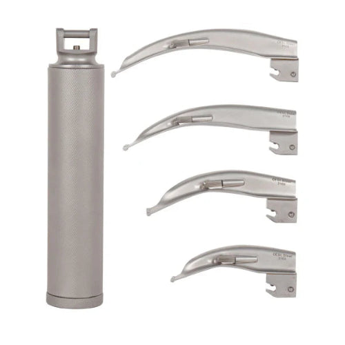
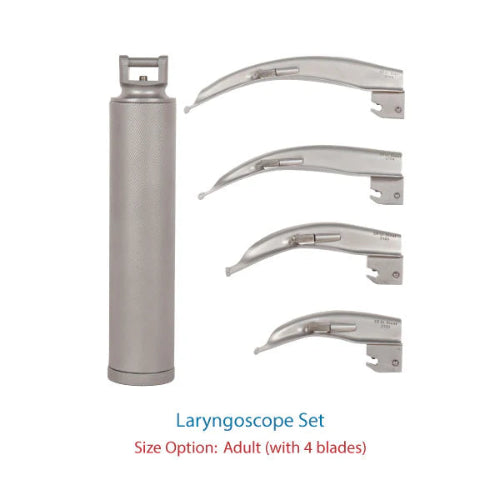
Laryngoscope Set Blade Size 2 3 and 4 Adult Macintosh Type


Type: Anesthesia
Laryngoscope Fiber Optic LED Set Macintosh 1-4 Blades Kit


Type: Scopes
Laryngoscope Premium with Blade Size 1, 2, 3, 4
Type: Scopes
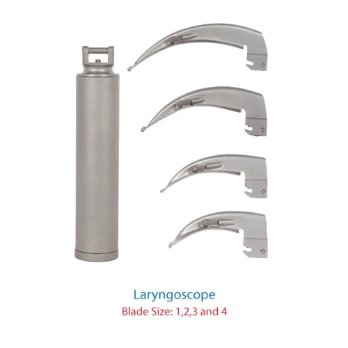
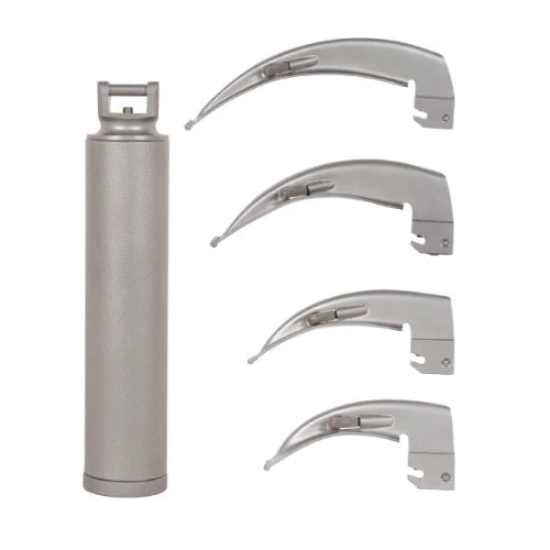
Laryngoscope Economy Blade Size 1, 2, 3, 4


Type: Scopes
Laryngoscope Premium with Blade Size 0, 1 in Pouch
Type: Scopes
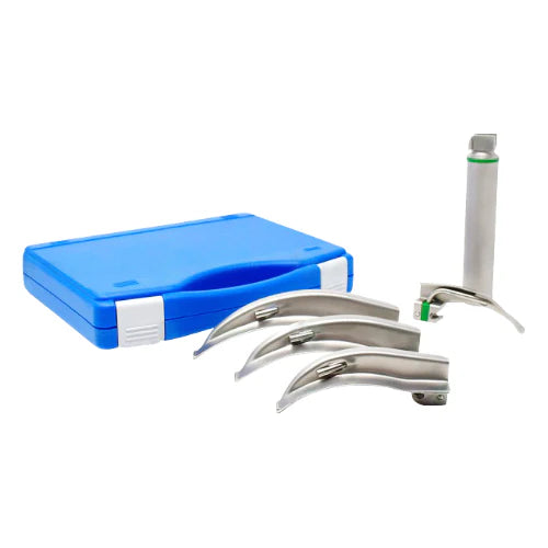
Fiber Optic LED Laryngoscope Set Macintosh 00,0,1


Type: Anesthesia
Reusable Portable Video Laryngoscope | 3 Blades


Type: Medansh
Portable Video Laryngoscope

Type: Scopes
Fiber Optic LED Laryngoscope Set Miller 00, 0, 1
Type: Scopes
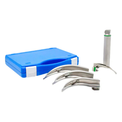
Fiber Optic LED Laryngoscope Set Macintosh 0,1, 2

Type: Anesthesia
Video Laryngoscpe with 2 Blades Disposable

Type: Medansh
Medical Portable Video Laryngoscope


Type: Medansh
Laryngoscope Premium with Blade Size 2, 3, 4

Type: Scopes
Fiber Optic LED Laryngoscope Set Miller 1, 2, 3

Type: Scopes
Fiber Optic LED Laryngoscope Set Miller 0, 1, 2
Collection:
Laryngoscope Maintenance: Best Practices Explained
You've seen it a hundred times in medical dramas: a patient is rushed into the emergency room, and a doctor inserts a metal tool into their mouth and says, "I'm in." That shiny device, which often looks like a futuristic handle with a curved blade, isn't for cutting or surgery. Its real job is much simpler and is one of the most important acts in modern medicine.
That critical tool is called a laryngoscope. Despite its intimidating appearance, it has only two primary functions.
- First, it acts like a specialized lever to gently move the tongue out of the way.
- Second, it shines a bright light toward the back of the throat, illuminating a path that would otherwise be completely hidden from view.
So, what is a laryngoscope used for , exactly? In practice, it's a guide. Imagine trying to see the bottom of a dark, cluttered bag; you'd need something to hold items aside and a light to see. The laryngoscope does just that for the airway, giving doctors a clear view of the windpipe. This simple-sounding procedure, known as laryngoscopy, is the crucial first step to safely placing a breathing tube.
This guide demystifies the device, transforming on-screen drama or pre-surgery anxiety into a clear process. It breaks down how the laryngoscope works and why it's a cornerstone of patient safety, one simple step at a time.
Why Doctors Can't Just "Look Down Your Throat"
If a patient needs help breathing, it seems logical that a doctor could just shine a light into their mouth to see the opening of the windpipe. In reality, it's not that simple. When a person is unconscious, their muscles---including the tongue---relax completely. The tongue, being a large muscle, often falls back and acts like a curtain, blocking any direct view into the back of the throat.
Deeper inside, there's another clever piece of anatomy at work: a small, leaf-shaped flap called the epiglottis. Think of it as a protective trapdoor that sits over your windpipe. Its everyday job is to close automatically when you swallow, keeping food out of your lungs. But in an unconscious patient, this trapdoor can cover the very opening the doctor needs to access.
This creates a two-part problem: the tongue is blocking the entrance, and the epiglottis is covering the destination. A simple flashlight can't physically move these obstacles. This is precisely what a laryngoscope is used for; a doctor needs a special tool that can gently manage the tongue, lift the epiglottis, and light up the path all at once.
Meet the Laryngoscope: The Key and Flashlight for the Airway
So, how do doctors get past the tongue and the epiglottis to see the airway? They use a purpose-built device called a laryngoscope. The easiest way to understand what a laryngoscope is used for is to think of it as a special key and a powerful flashlight combined into a single tool. It's designed not just to look into the throat, but to physically create a clear path to the destination.
The "key" function comes from the instrument's carefully shaped blade, which allows a doctor to gently sweep the tongue to the side and lift the epiglottis---that little trapdoor over the windpipe. This single, controlled motion opens up the view. At the same time, the "flashlight" function---a bright light located right near the tip---illuminates the way forward, revealing the entrance to the windpipe that would otherwise be hidden in shadow.
The laryngoscope's job is temporary. Its only purpose is to clear the path and light the way so a doctor can safely place a separate breathing tube. The laryngoscope is removed as soon as the breathing tube is secure. To accomplish this elegant task, the device itself is surprisingly straightforward, typically made of just three simple parts.
The Three Simple Parts of Every Laryngoscope
Despite its critical role, a laryngoscope is not a complex machine. It's built from three distinct pieces that work together in a simple, elegant system.
- The first and most obvious part is the handle. Think of it as the body of a sturdy flashlight---it's what the clinician holds, and it houses the batteries that power the device. Its weighted, textured grip gives the user precise control during the procedure.
- Attached to the handle is the most specialized component: the blade. This is the part that actually enters the mouth. While it might look intimidating, its job is to act like a carefully shaped, strong spoon, designed to gently sweep the tongue aside and lift anatomy out of the way. Blades are smooth, rounded, and come in different sizes to safely fit a patient's unique anatomy, from a newborn to a large adult.
- Of course, creating a path isn't helpful if you can't see where you're going. That's why every laryngoscope blade has a built-in light source. Located right near the tip of the blade, a tiny bulb or a fiber-optic channel projects a bright, focused beam of light exactly where it's needed, illuminating the entrance to the windpipe.
When a doctor prepares to use the tool, they click a sterile, single-use blade onto the handle. This simple connection completes an electrical circuit, instantly turning on the light. The three parts become one functional instrument, ready to create that clear, illuminated path to the airway.

Curved vs. Straight: Why Laryngoscope Blades Come in Two Main Flavors
Just as a mechanic needs more than one type of wrench, a doctor needs more than one type of laryngoscope blade. While they all perform the same job of moving the tongue and providing light, they come in two main "flavors": curved and straight. This variety isn't for show; it allows the clinician to choose the perfect tool to match a patient's unique anatomy, ensuring the safest and clearest view of the airway possible. The shape of the blade determines exactly how it creates that path.
- The most common design is the curved blade. Its gentle, C-shaped slope is designed to work in harmony with the natural curve of the tongue. The tip of this blade is placed in a small groove at the base of the tongue, just before the epiglottis. When the doctor gently lifts, the pressure on this spot indirectly pulls the epiglottis up and out of the way, revealing the entrance to the windpipe below. It's an elegant technique that uses the body's own structures to clear the view.
- In other situations, a straight blade offers a better solution. Unlike its curved counterpart, this thin, flat blade is designed to take a more direct approach. The doctor guides it past the tongue and slides it directly underneath the epiglottis. A gentle lift then raises the flap like a small garage door, providing an immediate, straight-line view to the airway. The choice between curved and straight is tailored to each individual, ensuring that no matter the situation, there is a tool designed to get the job done safely.
How Do You Assemble a Laryngoscope? It's Simpler Than You Think
With the correct blade chosen, the next step is connecting it to the handle. In a high-stakes medical situation, there's no time for complicated assembly with tiny screws or confusing instructions. Fortunately, the laryngoscope's design is brilliantly simple, engineered for speed and reliability when every second counts. This ensures the tool can be ready to use in a single, fluid motion.
Attaching the blade is a quick, two-part action.
- The base of the blade has a U-shaped hinge that hooks over a small bar on the handle, much like latching a sturdy gate.
- From there, the user swings the blade upward until it locks into a 90-degree angle with a firm click.
This single motion accomplishes two jobs at once: it secures the blade and, just as importantly, completes an electrical circuit, instantly turning on the light at the blade's tip.
This ingenious design means the device is ready for action the instant it's assembled---no fumbling for a separate light switch in a critical moment. That bright, steady light is essential for giving the doctor a direct line of sight down the patient's throat to the airway. But what if there was an even better way to see, without having to find the perfect angle? Modern technology has provided a powerful answer.
The Modern Upgrade: From Looking Directly to Watching on a Screen
That powerful answer is the video laryngoscope. To understand its benefit, think of the difference between parking a car using only your mirrors versus using a backup camera. A traditional laryngoscope is like using mirrors---it requires the doctor to get in just the right position to see the target directly. A video laryngoscope, however, is a game-changer that works more like that backup camera, giving the provider a perfect view every time.
Instead of the doctor having to crane their neck to get a direct line of sight, a video laryngoscope has a miniature camera built into the tip of the blade. This camera captures a live, high-resolution video from inside the throat and sends it to a small color screen mounted right on the handle. The doctor simply watches the screen to guide the breathing tube into place.
This simple change has a profound impact. It transforms a procedure that depends on a tricky alignment of the doctor's eye, the patient's mouth, and the airway into one guided by a clear, magnified image. The "indirect view" provided by the screen means the doctor doesn't need to look down the throat at all; they can see everything they need right in front of them, brightly lit and enlarged.
Ultimately, the video laryngoscope represents a major leap forward in making airway management easier and safer. By providing a consistently clear view of the vocal cords and the entrance to the windpipe, it helps turn a potentially challenging task into a more predictable and controlled process. This technological assist gives medical professionals a powerful advantage, especially in situations where a direct view might be difficult to achieve.

Why Doctors Value the 'Backup Camera' View for Your Airway
While the backup camera analogy helps explain how a video laryngoscope works, its true value lies in why it's needed. Just as some parking spots are tighter or more angled than others, every person's anatomy is slightly different. For medical professionals, this means getting a clear, straight line-of-sight into the airway isn't always a simple task.
Sometimes, a person's unique throat or neck structure can make it challenging to see the vocal cords with a traditional laryngoscope. Clinicians refer to this as a "difficult airway." This isn't a sign of a problem with the patient, but rather a physical puzzle for the doctor to solve. This is where the video laryngoscope shines, turning a potential blind spot into a clear, illuminated path on a screen and dramatically increasing safety.
This improved view delivers three huge benefits, making the process of securing an airway smoother and more reliable:
- A Clearer View for Everyone: The screen allows the entire medical team, not just one doctor, to see what's happening and assist more effectively.
- Making Difficult Airways Safer: It provides a reliable solution for those anatomical variations, giving doctors a powerful tool to handle challenging situations with confidence.
- An Invaluable Teaching Tool: For doctors-in-training, the screen allows them to see exactly what an experienced clinician sees, transforming a procedure that was once hard to teach into a shared visual lesson.
In the end, using a video laryngoscope often means the procedure can be done more gently, more quickly, and with greater predictability. It's all about using technology to make a critical task as safe as possible. Now that the laryngoscope has cleared the way and provided a perfect view, what is its ultimate goal? It's to guide the placement of the breathing tube itself.
The Real Goal: A Laryngoscope's Role in Placing a Breathing Tube
Once the doctor has that crystal-clear view of the vocal cords, the laryngoscope's main job is nearly done. Its entire purpose is to serve as a guide for something even more important: a breathing tube. This soft, flexible tube (formally called an endotracheal tube) is what actually keeps you breathing safely. Think of the laryngoscope as a tool that helps you see the eye of a needle in a hard-to-reach spot; it doesn't do the sewing itself, but it makes threading the needle possible.
With the path illuminated and clear, the doctor gently guides the breathing tube past the vocal cords and into the windpipe. As soon as the tube is securely in place, the laryngoscope is carefully removed. This is a key point that often gets confused: the laryngoscope is only in the mouth for a very short time---typically less than a minute. It's the breathing tube that stays in place to deliver oxygen and anesthesia, acting as a secure and dedicated highway for air to get to the lungs.
This entire procedure, from using the laryngoscope to placing the tube, is called intubation. It is the gold standard for protecting a person's airway during surgery or a critical medical emergency. By creating this direct path for breathing, doctors ensure the body gets the oxygen it needs, allowing them to focus on the surgery or treatment at hand. But does a procedure this important count as surgery itself?
Is a Laryngoscopy Considered Surgery?
That's an excellent question, and the short answer is no---a laryngoscopy is considered a medical procedure, not a surgery. The simplest way to think about the difference is that surgery almost always involves cutting or making an incision to repair or remove tissue. A procedure, on the other hand, is a task performed to diagnose or treat a condition, and in this case, it doesn't involve any cutting at all. It's about viewing and guiding, not operating.
The key difference lies in the goal. As we've seen, when used for intubation before an operation, the laryngoscope is just a temporary guide. However, a laryngoscopy can also be a standalone diagnostic tool. For example, if you have a persistent cough or hoarseness, an Ear, Nose, and Throat (ENT) specialist might perform a laryngoscopy in their office to get a clear, magnified look at your vocal cords and throat to find the cause of the problem.
So, a laryngoscopy can be either a quick step before surgery (for intubation) or a diagnostic examination all on its own. This distinction is important because the experience is very different depending on the purpose. Knowing it can be done for these different reasons naturally brings up a common concern: How painful is a laryngoscopy?
The Big Question: How Painful Is a Laryngoscopy?
It's completely natural to worry about discomfort when it comes to any medical procedure involving your throat. The answer to this question, however, depends entirely on the situation. The key is to remember that doctors have excellent methods to ensure you are comfortable, whether you are asleep for surgery or awake for a quick examination.
For the vast majority of cases seen in hospitals or medical dramas, a laryngoscopy is performed to place a breathing tube before surgery. In this scenario, you will be under general anesthesia, meaning you are completely asleep and unaware. You won't feel, see, or remember the procedure at all. The medical team only performs the laryngoscopy after you are fully anesthetized, guaranteeing a painless experience.
What about when you're awake, like in an Ear, Nose, and Throat (ENT) specialist's office? In this case, the doctor's top priority is to prevent pain and suppress your natural gag reflex. They do this by applying a topical anesthetic, usually a numbing spray or gel, to the back of your throat. It's a similar feeling to the numbing gel a dentist uses on your gums before giving an injection---the area quickly feels thick and numb.
Because of this effective numbing, most people do not describe an awake laryngoscopy as painful. Instead, they report an odd sensation of pressure or a feeling that "something is there," but without any sharpness. The goal is always to make the brief examination as tolerable as possible, and these numbing agents are extremely effective at achieving that.
What to Expect After: Can I Eat and Talk Normally?
Once the procedure is over and any numbing medication has worn off, your main questions will likely be about getting back to normal. The laryngoscopy recovery period is typically very short, but you can expect a few temporary changes. The most common after-effect is a mild sore throat, often feeling similar to the very beginning of a cold. Your voice might also be a little hoarse or raspy for a short while.
This happens simply because a medical instrument, no matter how gently used, can cause minor irritation to the sensitive tissues in your throat. Think of it like the slight muscle soreness you might feel the day after trying a new exercise---it's a sign that the area was worked, and it fades quickly. For most people, any soreness or hoarseness is gone within a day or two.
So, can you eat after a laryngoscopy? Yes, but you can make yourself more comfortable by being mindful of your choices for the first day. To soothe any irritation, focus on simple remedies:
- Sip cool liquids like water or suck on ice chips.
- Eat soft foods like yogurt, pudding, or soup.
- Avoid scratchy or acidic foods like toast, chips, or citrus.
These small steps can make a big difference in your comfort. While a temporary sore throat is the most common and mildest of risks, it's helpful to understand the bigger picture of how doctors ensure the procedure is overwhelmingly safe.
How Doctors Keep Laryngoscopy Safe
It's natural to wonder about potential issues beyond a temporary sore throat. The primary concern for laryngoscopy, though very uncommon, involves the instrument accidentally chipping or damaging a tooth. To prevent this, doctors treat the procedure with incredible care, much like guiding a key into a delicate lock in the dark---the goal is precision, not force. Significant complications are rare, a direct result of both training and technology.
- A doctor's skill is the first and most important line of defense. Performing a laryngoscopy is a refined technique that medical professionals practice extensively until it becomes second nature. They learn to use the laryngoscope as a tool of finesse, gently lifting the tongue and tissues to create a clear line of sight, rather than pushing through them. This emphasis on gentle technique is the main reason the procedure is overwhelmingly safe when performed by a trained professional.
- On top of that expertise, modern tools have made the process even safer. Many hospitals now use video laryngoscopes, which feature a tiny camera on the tip of the blade. This works just like a car's backup camera, sending a clear, magnified picture to a monitor. This enhanced view allows the doctor to see exactly where the instrument is, enabling more precise movements and dramatically reducing the chance of accidental contact with teeth or other tissues. This combination of skill and technology ensures patient safety is the top priority.
The Experts Behind the Scope: Who Performs a Laryngoscopy?
So, who is the expert wielding this important tool? In surgery or an emergency, it's most often an anesthesiologist or a critical care doctor. Their goal is to secure a breathing airway for an unconscious patient. They use the rigid laryngoscope to clear a path for a breathing tube, ensuring the patient receives oxygen safely during a procedure or critical illness.
Laryngoscopy isn't just for anesthesia, though. If you have a persistent hoarse voice, you might see an Ear, Nose, and Throat specialist (ENT). ENTs perform laryngoscopies for diagnosis---to get a close-up look at your vocal cords and surrounding tissues to find the cause of your symptoms, rather than to place a breathing tube.
To do this while a patient is awake, ENTs use a different tool: a flexible laryngoscope. Instead of a rigid metal blade, this is a thin, pliable tube with a camera, much like a strand of cooked spaghetti. Passed gently through the nose, it navigates the throat's natural curves to get a clear view without needing a large, rigid instrument.
This explains the two very different experiences. Intubation laryngoscopy involves a rigid scope while you're fully asleep. A diagnostic laryngoscopy, however, uses a flexible scope while you're awake in an ENT's office. A numbing spray keeps you comfortable as the doctor watches your vocal cords move, which provides vital diagnostic clues.
A Deeper Look: Direct Laryngoscopy and Biopsy
While ENTs often use a flexible scope in their office, sometimes they need a more stable, hands-on approach for a closer look. This is where a Direct Laryngoscopy (DL) comes in. As the name implies, the ENT specialist uses a rigid laryngoscope to get a straight-line, direct view down the throat to the voice box. Think of it like a mechanic using a specialized tool to look directly into a hard-to-reach part of an engine, instead of just using a camera on a wire. This method provides an unobstructed and stable view for performing delicate tasks.
Often, the main reason for a Direct Laryngoscopy is to perform a biopsy. If the specialist sees an unusual growth or patch of tissue on the vocal cords, a visual inspection isn't enough to make a diagnosis. A biopsy is simply the process of taking a tiny tissue sample---often no bigger than the tip of a pen---from that specific area. Just as a gardener might snip a small piece of a leaf to identify a plant disease, this tissue sample is sent to a laboratory to be examined under a microscope, which is the only way to get a definitive answer.
So, is a laryngoscopy with a biopsy a surgery? Yes, it's considered a minor surgical procedure. Because it requires you to be perfectly still and comfortable, it's performed in an operating room under general anesthesia. This allows the ENT to work precisely without causing any discomfort. Combining the direct view with the ability to take a sample makes this procedure one of the most effective ways for doctors to diagnose and ultimately treat issues deep within the throat.
From Mystery Tool to Lifesaving Essential
What may have once seemed like a complex or intimidating tool in a fast-paced medical drama is now clear. The laryngoscope is a brilliantly simple solution to a fundamental challenge---how to get a clear, safe view of the airway when the tongue and other tissues are in the way.
At its heart, the device performs two elegant tasks: it acts as a gentle lever to move anatomy aside, and it shines a light to illuminate the path forward. This straightforward laryngoscope usage is the critical first step that enables a clinician to safely guide a breathing tube, ensuring a patient can breathe with support, whether it's with a traditional blade or a modern video screen.
With this knowledge, you are now equipped to see things differently. The next time you watch a medical show or hear a loved one talk about an upcoming surgery, you can confidently understand the crucial role of laryngoscopy in anesthesia. You won't just see a tool; you'll recognize the purpose behind it and appreciate the deliberate, careful steps taken to protect a patient.
Ultimately, the laryngoscope is more than just a piece of metal and light. It is a bridge between a problem and its solution, a key that unlocks a safe passage for breath. This knowledge transforms it from an object of anxiety into a powerful symbol of modern medicine's quiet, constant commitment to patient care.
Q&A
Question: What exactly does a laryngoscope do?
Short answer: It gently moves the tongue out of the way and shines a bright light to reveal the entrance to the windpipe. In practice, the laryngoscope acts like a specialized lever and flashlight combined: its blade sweeps the tongue aside and helps lift the epiglottis so clinicians can see the airway. Its role is temporary—once it provides a clear view, it guides the placement of a breathing tube (endotracheal tube) during intubation and is then removed, typically within a minute.
Question: Why can’t doctors just look down the throat with a flashlight?
Short answer: In an unconscious person, the relaxed tongue falls back and blocks the view, and the epiglottis can cover the windpipe like a trapdoor. A simple light can’t move these structures. A laryngoscope is specifically shaped to gently manage the tongue, lift the epiglottis, and illuminate the path simultaneously, creating the clear line of sight a flashlight alone can’t provide.
Question: What’s the difference between curved and straight laryngoscope blades?
Short answer: Both clear a path to the airway, but they do it differently to suit different anatomies. A curved blade follows the tongue’s natural curve; its tip sits in the groove at the base of the tongue and, when lifted, indirectly raises the epiglottis to reveal the windpipe. A straight blade takes a direct approach: it slides under the epiglottis and lifts it like a small door, providing a straight-line view. Clinicians choose the shape that gives the safest, clearest view for each patient.
Question: What is a video laryngoscope and why do clinicians value it?
Short answer: It’s a laryngoscope with a tiny camera at the blade tip that displays a live, magnified view on a screen—like a backup camera for the airway. This “indirect view” reduces the need for perfect eye–mouth–airway alignment, often making the procedure gentler, quicker, and more predictable. It’s especially helpful for difficult airways, lets the whole team see and assist, and is an excellent teaching tool.
Question: Will a laryngoscopy hurt, and what should I expect afterward?
Short answer: For intubation before surgery, you’re under general anesthesia—you won’t feel or remember it. For an awake diagnostic exam (often with an ENT), a numbing spray or gel is used; most people feel pressure or a foreign-body sensation rather than pain. Afterward, a mild sore throat or brief hoarseness is common and usually resolves within a day or two. Comfort tips for the first day include sipping cool liquids, choosing soft foods (yogurt, soup), and avoiding scratchy or acidic items.