Type: Scopes
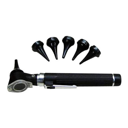
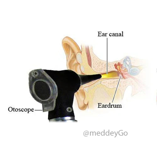
Basic Student Pocket Scope, Otoscope & Auriscope
Collection:
Microscope Techniques: Enhance Your Research
Microscope Techniques for Advanced Research
In the intricate world of scientific research, the microscope stands as an indispensable tool. Its ability to magnify the minutest details allows researchers to explore the unseen realms of life and matter. Whether you're delving into biological cells or inspecting electronic components, understanding the various microscope techniques and types available will enhance your research capabilities. By delving deeper into these powerful instruments, researchers can unlock new insights and push the boundaries of science further than ever before.
Summary
This guide surveys major microscope types (compound, dissecting/stereo, digital, electron, portable), core performance factors (resolving power, lens quality, ergonomics, accessories), and advanced techniques (fluorescence, confocal, phase contrast, polarized light). It connects capabilities to applications across biology, materials science, electronics, and forensics, showing how technique choice drives insight. Practical selection advice emphasizes aligning purpose, features, and budget, supported by brand research and reviews. A closing Q&A addresses common decisions about magnification, technique selection, and when ultra-high-resolution electron microscopy is warranted.

Types of Microscopes
Microscopes are diverse, and selecting the right one depends on your specific research requirements.
- Compound Microscope: This is the most common type used in laboratories. It employs multiple lenses to achieve high magnification, making it ideal for viewing small specimens like bacteria or thin tissue slices. The price can vary significantly based on the brand and features, but they generally offer excellent value for educational and basic research purposes. Compound microscopes are particularly favored for their versatility and ability to provide detailed views of cellular structures.
- Dissecting Microscope: Also known as a stereo microscope, it provides a three-dimensional view of larger specimens. This type is perfect for dissection or examining objects like rocks, insects, and plants. The stereo view offered by these microscopes makes them invaluable for tasks requiring depth perception and precise manipulation, such as in botany or entomology.
- Digital Microscope: Equipped with a camera, this microscope connects to a computer or a screen, allowing for easy observation and sharing of images. It's particularly useful for presentations and detailed image analysis. The ability to capture and digitally manipulate images makes digital microscopes a favorite in educational settings and collaborative research environments.
- Electron Microscope: Offering the highest resolving power of all microscopes, electron microscopes use beams of electrons instead of light. They can magnify specimens up to two million times, making them essential for advanced research. However, the electron microscope price can be prohibitive, limiting their use to specialized institutions. Despite their cost, electron microscopes are indispensable for fields requiring ultra-high resolution, such as nanotechnology and molecular biology.
- Portable Microscope: Compact and lightweight, these microscopes are designed for fieldwork. They are often used for environmental studies and mobile phone repairs. Their portability makes them ideal for researchers who need to conduct analyses on-site, providing a practical solution for field biologists and technicians alike.
Key Features and Accessories
Selecting the right microscope involves understanding its key features and accessories:
- Resolving Power: This determines the microscope's ability to distinguish two close points as separate entities. A higher resolving power indicates better clarity and detail. This is particularly important in disciplines where fine structural details are critical, such as pathology or materials science.
- Microscope Lens: The quality of the lenses affects the image clarity. Opt for lenses with high numerical apertures for better resolution. Investing in high-quality optics can dramatically improve the performance of a microscope, offering clearer and more accurate images.
- Binocular Microscopes: These have two eyepieces, reducing eye strain and providing a more comfortable viewing experience during prolonged use. For researchers spending long hours at the microscope, this feature can significantly enhance comfort and productivity.
- Accessories: Items such as slides, coverslips, and illumination sources enhance the functionality of a microscope. Specialized accessories like phase contrast and darkfield attachments can also be used to improve image quality. These tools allow researchers to tailor the microscope's capabilities to their specific needs, expanding the range of potential applications.
Advanced Microscope Techniques

For those engaged in advanced research, mastering specific microscope techniques is crucial. These techniques not only enhance the quality of observations but also expand the range of research applications. By employing advanced methods, researchers can delve deeper into the microcosm, uncovering new insights that are not possible with basic techniques alone.
Fluorescence Microscopy
This technique uses fluorescent dyes to stain specific components of a specimen, making them glow under ultraviolet light. It's particularly useful in cellular and molecular biology for identifying proteins, nucleic acids, and other vital molecules within cells.
Fluorescence microscopy allows for the visualization of specific structures within complex biological systems. By tagging different cellular components with distinct fluorescent markers, researchers can observe interactions and processes in real-time. This technique is indispensable in the study of cellular dynamics and molecular pathways, providing insights into cell function and disease mechanisms.
Confocal Microscopy
Confocal microscopy employs a laser to illuminate a specimen and a pinhole to eliminate out-of-focus light. The result is a clear, detailed image with depth, allowing for the reconstruction of three-dimensional structures from the collected data.
This technique is essential for examining thick specimens where detail at various depths is required. By capturing multiple images at different focal planes, confocal microscopy can create comprehensive 3D models of structures, making it invaluable in fields such as neurobiology and developmental biology. Additionally, the precision of confocal microscopy enables researchers to study cellular processes in living specimens with minimal interference.
Phase Contrast Microscopy
Phase contrast microscopy is designed to enhance the contrast in transparent specimens without the need for staining. It's particularly beneficial for observing living cells and tissues in their natural state.
This technique is advantageous for researchers who require non-invasive observation methods. By improving contrast in otherwise transparent samples, phase contrast microscopy reveals details that are often invisible with standard brightfield microscopy. This makes it an essential tool in live cell imaging, facilitating studies in cell biology and microbiology without altering the specimen.
Polarized Light Microscopy
This technique is used to study materials that exhibit birefringence, such as crystals and fibers. By analyzing how these materials affect polarized light, researchers can gain insight into their structure and composition.
Polarized light microscopy is crucial in geology and material science, where understanding the crystalline structure of minerals and synthetic materials is vital. By revealing stress patterns and structural orientation, this technique helps in assessing material properties and behavior under different conditions.
Practical Applications of Microscopes

Microscopes find applications across various fields, from biology to electronics, each benefiting from the enhanced observation capabilities these instruments provide. The versatility of microscopes makes them indispensable tools in both research and applied sciences, transforming the way we understand and manipulate the world at a microscopic level.
Biological Research
In biology, microscopes are used to study cells, tissues, and microorganisms. Techniques like fluorescence microscopy have advanced cancer research, while electron microscopes have provided insights into the structure of viruses.
Biological research relies heavily on microscopes to uncover the mysteries of life. From understanding cellular functions to exploring genetic material, microscopes enable breakthroughs in medical research and biotechnology. By allowing scientists to visualize the intricacies of biological systems, these tools have accelerated advancements in drug development, genetic engineering, and disease diagnostics.
Material Science
Material scientists use microscopes to analyze the structural properties of metals, ceramics, and polymers. Electron and polarized light microscopes help reveal details that are crucial for developing new materials.
In material science, microscopes are essential for exploring the microstructure and properties of materials. By examining surfaces, interfaces, and defects at high magnification, researchers can develop materials with enhanced performance and durability. This knowledge is vital for innovations in engineering, nanotechnology, and sustainable material design.
Electronics and Mobile Repair
In the realm of electronics, microscopes are essential for inspecting and repairing small components. Portable and digital microscopes are commonly used in mobile phone repair, allowing technicians to see tiny circuits and connections clearly.
Microscopes are critical in the electronics industry, where precision is key. They allow for the detailed inspection and repair of intricate electronic components, ensuring quality and functionality. As devices become increasingly miniaturized, the role of microscopes in electronics continues to grow, supporting advancements in technology and manufacturing.
Forensics
In forensic science, microscopes play a vital role in analyzing evidence such as hair, fibers, and other trace materials. The ability to examine these details can be critical in solving criminal cases.
Forensic microscopes provide the detailed analysis necessary for evidence evaluation. By examining microscopic details, forensic scientists can link suspects to crime scenes, identify substances, and uncover hidden evidence. This precision is crucial for building cases and ensuring justice in the legal system.

Choosing the Best Microscope
Selecting the best microscope for your needs requires careful consideration of your research objectives and budget constraints. Here are a few tips:
Determine Your Purpose
Understand what you need the microscope for. Is it for educational purposes, advanced research, or practical applications like mobile repair? Knowing your primary use will guide your selection process, ensuring that you choose a microscope that meets your specific needs and enhances your research capabilities.
Consider the Type
Based on your purpose, choose the type of microscope that offers the features you need. For detailed cellular studies, a digital or electron microscope might be necessary, while a dissecting microscope is ideal for larger specimens. Each type of microscope offers unique advantages, so aligning your choice with your research goals is essential for maximizing effectiveness.
Budget Wisely
While high-end microscopes offer advanced features, they come at a higher cost. Balance your needs with your budget, and consider starting with a basic model that can be upgraded with accessories over time. Investing strategically allows you to access the essential features you need while leaving room for future enhancements as your research progresses.
Research Brands and Reviews
Look for reputable brands and read reviews to ensure the microscope you choose is reliable and of high quality. Customer reviews and expert opinions provide valuable insights into the performance and durability of different models, helping you make an informed decision.
Conclusion
Microscopes are powerful tools that have revolutionized research across numerous fields. By understanding the different types, techniques, and practical applications, you can enhance your research capabilities and achieve more accurate and detailed observations. Whether you're a seasoned researcher or just starting, investing in the right microscope will undoubtedly enrich your scientific endeavors. With the right tools and knowledge, the microscopic world reveals endless opportunities for discovery and innovation.
Frequently Asked Questions
Question: How do I choose between a compound, dissecting, digital, electron, and portable microscope? Short answer: Match the microscope to your task. Use a compound microscope for high-magnification views of small, thin specimens like bacteria or tissue slices. Choose a dissecting (stereo) microscope for a three-dimensional view of larger objects (e.g., insects, plants, rocks) and tasks needing depth perception. Pick a digital microscope when you need to capture, display, or share images easily for presentations or collaborative analysis. Opt for an electron microscope when ultra-high resolution is essential (e.g., nanotechnology, molecular biology), recognizing that high cost often limits access to specialized institutions. Select a portable microscope for on-site work in the field or precision tasks like mobile phone repair. Start with the type that fits your primary purpose and budget, then expand with accessories as needs grow.
Question: Does higher magnification always mean a clearer image? Short answer: No. Clarity depends more on resolving power and lens quality than on magnification alone. Resolving power determines how well two close points can be distinguished; higher resolving power yields more detail. High-quality lenses with greater numerical apertures improve resolution, and good optics generally enhance image accuracy. Comfort features like binocular eyepieces reduce eye strain for long sessions, and appropriate accessories (e.g., illumination, contrast methods) can further improve image quality.
Question: When should I use fluorescence microscopy versus confocal microscopy? Short answer: Use fluorescence microscopy when you need to label and visualize specific molecules or structures (e.g., proteins, nucleic acids) within cells, often to observe interactions and processes in real time. Choose confocal microscopy when you need optically crisp images from thick specimens and the ability to reconstruct three-dimensional structures by collecting images at multiple focal planes. Confocal's laser illumination and pinhole improve detail and reduce out-of-focus light, and it enables study of cellular processes in living specimens with minimal interference.
Question: What's the best technique for observing living, unstained cells? Short answer: Phase contrast microscopy. It enhances contrast in transparent, unstained specimens, revealing details that standard brightfield often misses. This makes it ideal for non-invasive, live-cell observations in cell biology and microbiology. If depth-resolved imaging of living specimens is needed, confocal microscopy can also be useful while minimizing interference, though it is typically applied for detailed optical sectioning.
Question: Are electron microscopes worth the price, and who should consider them? Short answer: They are worth it when your work demands the highest resolving power and extreme magnification---up to about two million times---such as in nanotechnology, molecular biology, or detailed materials analysis. Because their cost is prohibitive, they are usually housed in specialized institutions. If your questions can be answered with light-based methods (compound, fluorescence, confocal, polarized light), those are more accessible; move to electron microscopy only when ultra-high resolution is essential.
Question: What's the difference between magnification, resolving power, and contrast---and which matters most for image quality? Short answer: Magnification makes the image larger, resolving power determines how well you can distinguish two close points as separate, and contrast is how clearly features stand out from the background. For useful, clear images, resolving power and contrast matter more than magnification alone. High-quality lenses with greater numerical apertures improve resolution, while appropriate accessories (e.g., illumination, phase contrast, darkfield attachments) enhance contrast. Comfort features like binocular eyepieces also help sustain image quality over long sessions by reducing eye strain.
Question: When is polarized light microscopy the right choice, and what can it reveal?
Short answer: Use polarized light microscopy for birefringent materials such as crystals and fibers---common in geology and materials science. By analyzing how a sample alters polarized light, you can uncover structural orientation, crystalline organization, and stress patterns. These insights help assess composition and behavior of minerals and synthetic materials under different conditions.
Question: How do digital microscopes improve teaching, documentation, and collaboration?
Short answer: Digital microscopes integrate a camera and connect to a computer or display, making it easy to view, capture, share, and analyze images. They're ideal for presentations, collaborative review, and detailed image analysis. The ability to record and manipulate images supports education, team-based research, and streamlined documentation.
Question: I'm on a tight budget---how should I choose and upgrade a microscope over time?
Short answer: Start by defining your primary purpose (e.g., education, advanced research, fieldwork, electronics repair) and pick the type that fits those needs. Balance must‑have features with cost, then plan to expand capabilities with accessories (better illumination, phase contrast or darkfield attachments, quality slides and coverslips) as your work evolves. Research reputable brands and read customer/expert reviews to ensure reliability and durability before you buy.
Question: Which microscopes are best for electronics inspection and mobile phone repair?
Short answer: Portable and digital microscopes are commonly used for electronics work. Portables are compact and field‑ready for on‑site inspections, while digital microscopes let you clearly view tiny circuits and connections on a screen, capture images for documentation, and collaborate during diagnostics and repair.
Question: How do I match my research application to the right microscope or technique?
Short answer: Start with your goal, then pick the tool that reveals the needed detail. For cellular work, use a compound microscope; add fluorescence to label specific molecules and confocal to get crisp optical sections and 3D reconstructions in thick samples. For living, transparent cells that you don’t want to stain, choose phase contrast. For materials and geology, use polarized light microscopy to study birefringent crystals and fibers, and consider electron microscopy when ultra‑high resolution is essential. For electronics inspection and mobile repair, portable and digital microscopes make on‑screen viewing and fieldwork practical. In forensics, dissecting (stereo) and compound microscopes help analyze hair, fibers, and trace materials, with polarized light aiding structure assessment.
Question: I need three-dimensional information—should I choose a stereo (dissecting) microscope or confocal microscopy?
Short answer: Choose a stereo microscope for larger, opaque specimens when you need real-time depth perception and precise manipulation (e.g., insects, plants, rocks). Choose confocal microscopy when you need depth-resolved, high‑detail imaging at the cellular level, especially in thick or complex specimens. Confocal uses laser illumination and a pinhole to reject out‑of‑focus light, enabling sharp optical sections and 3D reconstructions.
Question: My images look dull or unclear—what features or accessories should I adjust first?
Short answer: Prioritize resolution, contrast, and illumination. Use high‑quality lenses with greater numerical apertures to improve resolving power. Optimize illumination and consider contrast‑enhancing accessories such as phase contrast or darkfield attachments. Basic essentials like proper slides and coverslips also matter. For comfort and consistency during long sessions, binocular eyepieces reduce eye strain and help you maintain image quality.
Question: Should I stain with fluorescence or keep specimens unstained with phase contrast for live-cell work?
Short answer: Use phase contrast when you want to observe transparent, living cells in their natural state without staining—it boosts contrast without altering the specimen. Use fluorescence when you need molecular specificity, such as highlighting proteins or nucleic acids and tracking their interactions in real time. If you also need depth-resolved views in thicker samples, confocal microscopy can provide crisp optical sections while supporting live imaging with minimal interference.