Type: Scopes
Direct Ophthalmoscope Set Delux Quality
Type: Scopes
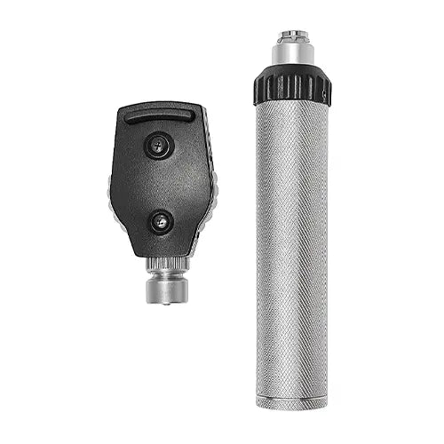
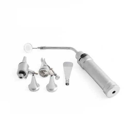
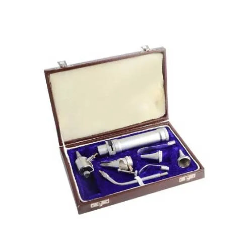
Direct Oto-Opthalmoscope Set
Type: Scopes
ENT Hein Type Set Premium Quality
Type: Scopes
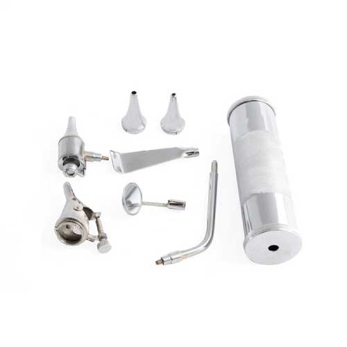
ENT Diagnostic Set with Brass Handle
Type: Scopes
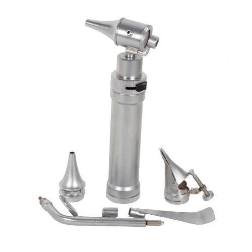
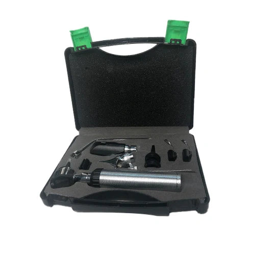
ENT Diagnostic Set with Small Torch
Type: Scopes
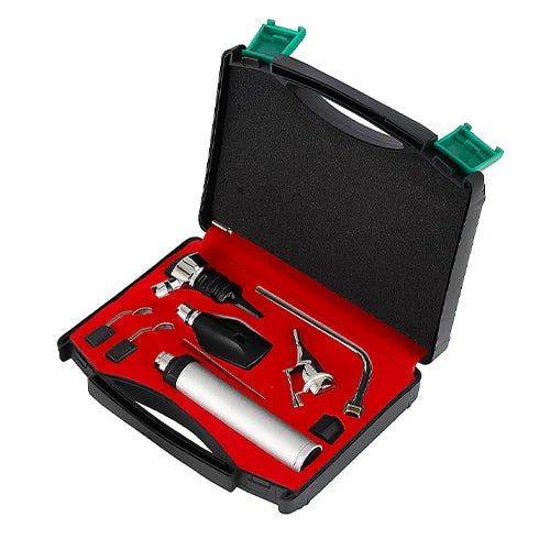
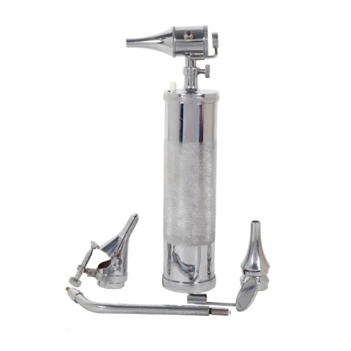
ENT Diagnostic Complete Set with Opthalmoscope Otoscope


Type: Scopes
Ophthalmoscope Imported Premium for Eye Exam
Collection:
How to Use an Ophthalmoscope Effectively
You know the moment. The lights dim, the doctor leans in with that familiar instrument, and a surprisingly bright light shines right into your eye. You try your best not to blink, wondering what on earth they are actually looking for in there. For many, it's a slightly awkward part of a check-up, but the answer to that question is far more important than just checking your vision.
That little device, officially called an ophthalmoscope, is much more than just a lighted magnifying glass. While it is an essential eye examination tool, its real power lies in providing a direct, real-time look at your nerves and blood vessels. In practice, it turns your eye into a unique window, offering a view into your body's overall health that a doctor can't get anywhere else without invasive procedures.
Because the eye offers this clear view, this simple, painless test can reveal the earliest signs of systemic health problems. Subtle changes in the delicate blood vessels at the back of your eye can signal developing conditions like diabetes, high blood pressure, or even high cholesterol, often long before other symptoms appear.
Summary
An ophthalmoscope provides a direct, painless view of the retina, optic nerve, and retinal vessels, offering insights into both eye and overall health. Clinicians use direct scopes for detailed close-ups and indirect scopes for wide retinal surveys, check the red reflex, and often dilate pupils for a comprehensive exam. Retinal changes can reveal early signs of diabetes, hypertension, and raised intracranial pressure before other symptoms emerge. Knowing what the tool shows—and how it differs from similar instruments—helps you engage confidently in this quick, potentially life‑saving assessment.
What Is That "Eye-Light Thingy" and Why Does It Matter?
An ophthalmoscope is a powerful, lighted magnifying glass designed specifically to let your doctor peer through your pupil and see all the way to the back of your eye. They are looking at the retina---the thin layer of tissue at the very back of your eyeball that acts like the sensor in a digital camera.
This instrument also provides a clear picture of the delicate blood vessels and the optic nerve, which is the main cable connecting your eye to your brain. Your eye is the only place on your body where a doctor can directly see blood vessels in their natural state, without any cutting. What eye doctors see with their light can reveal the earliest signs of systemic conditions, making this simple check incredibly important for more than just your vision.
Direct vs. Indirect: The Two Types of Ophthalmoscopes You'll Encounter
Depending on the reason for your visit, the ophthalmoscope your doctor uses might look different. The most common version, often seen during a routine physical, is a small, handheld device called a direct ophthalmoscope. Think of it as a powerful, self-lit magnifying glass. It gives your doctor a highly detailed, up-close view of specific parts of your retina, like the optic nerve head or a single blood vessel. This is perfect for a quick, targeted health screening.
You may also have seen an eye specialist wear a more complex-looking device that resembles a headlamp combined with a pair of glasses. This is the indirect ophthalmoscope. While it looks more intimidating, its purpose is to give your doctor a much wider, panoramic view of the retina. Instead of peeking through a keyhole (the direct ophthalmoscope), the doctor can now see the whole room at once.

The choice between the two types involves trading detail for scope. The handheld direct scope is like using binoculars to focus on a single bird in a tree---you see every feather in stunning detail. The headset-style indirect scope is like taking a wide-angle photograph of the entire forest---you might not see the individual feathers, but you can check the health of all the trees at once.
Ultimately, the instrument depends on what your doctor needs to see. A quick check might only require the handheld device. However, for a comprehensive exam, especially if your pupils are dilated, the doctor will likely use the indirect ophthalmoscope to get a complete picture and ensure no part of your retina is left unturned.
A Guided Tour Inside Your Eye: What Is the Doctor Actually Seeing?
When your doctor peers through the ophthalmoscope, they get a direct look at the very back of your eyeball. The main structure they see is your retina, a thin layer of tissue that acts like the sensor in a digital camera. This is where light entering your eye is turned into a picture. A healthy retina is crucial for clear vision, so the doctor checks to ensure this vital "screen" looks smooth and intact.
Just as important is how that picture gets to the brain. The doctor also examines your optic nerve, which appears as a small spot where it connects to the retina. Think of the optic nerve as the data cable that plugs your eye into your brain, sending all visual information for processing.
Together, these parts---along with the tiny blood vessels that nourish them---create a living map at the back of your eye. By looking at these structures, your doctor isn't just checking your vision; they're getting a unique snapshot of your body's well-being. This simple process is an incredibly effective way to spot problems early.
What a Healthy "Fundus" Looks Like (And Why It's Good News)
So, what is the doctor hoping to see? A healthy retina has a consistent, peachy-orange color, similar to the shade of a hazy sunset. Against this smooth background, your optic nerve stands out as a brighter, well-defined circle---the central hub where all the visual information prepares to leave the eye. Seeing this clear, uniform landscape is the first sign that everything is in good working order.
The doctor will also pay close attention to the delicate network of blood vessels that spread across your retina. Think of them as tiny, crisp red rivers flowing across the orange landscape. In a healthy eye, these vessels have sharp, clear edges. The doctor is checking to make sure they aren't blurry, swollen, or showing any signs of leaking, which helps confirm your retina is getting the steady blood supply it needs.
When your doctor pulls the ophthalmoscope away and tells you that "everything looks great back there," this is what they mean. They've seen a smooth retina, a healthy-looking optic nerve, and a clear map of blood vessels. One of the very first things they check for is a specific healthy glow known as the red reflex.
The "Red Reflex" Test: Why Your Doctor Wants to See Red
Have you ever taken a flash photograph and noticed someone's eyes glowing red in the picture? That familiar "red eye" effect is almost exactly what your doctor is looking for in one of the first parts of an eye exam. This reddish-orange glow, known as the red reflex, is the light from the ophthalmoscope bouncing off the blood-rich retina at the back of your eye. Seeing it is a very good sign.
The presence of a bright, even red reflex tells the doctor that the path of light is clear. Think of it like shining a flashlight down an empty hallway---the light hits the far wall and reflects back. If something were blocking the hallway, you wouldn't see the reflection. In the eye, a strong red reflex suggests that there are no major obstructions, like a dense cataract, clouding the view.
This simple test acts as a crucial first screening. Once the doctor confirms a healthy red reflex, they know they have a clear window to look deeper and examine the fine details of your retina and optic nerve, where more subtle clues about your overall health are often found.

How Your Eyes Can Reveal Early Signs of Diabetes
It might seem strange, but one of the first places diabetes can show up is inside your eye. This is because your eye is the only place in the entire body where a doctor can directly see your blood vessels in action without needing to perform surgery. That clear view, provided by the ophthalmoscope, acts as an incredible early warning system.
Diabetes involves high blood sugar, and over time, that excess sugar can damage the body's smallest blood vessels. Imagine a fragile garden hose that becomes brittle under too much pressure; eventually, it might start to develop tiny cracks and leaks. The same process can happen to the fine blood vessels that nourish your retina.
Using the ophthalmoscope, your doctor can get a highly magnified look at this network of vessels. They are searching for microscopic leaks or tiny spots of blood that signal the vessels are under stress. Discovering these subtle clues is one of the most important applications of the ophthalmoscope, often allowing for the detection of diabetic changes at their earliest stage. Remarkably, these changes can sometimes appear before a person experiences any other classic symptoms of diabetes, leading to an earlier diagnosis and treatment.
Spotting High Blood Pressure by Looking at Your Retina
Just as the eye's tiny vessels can reveal signs of high blood sugar, they also act as a pressure gauge for your cardiovascular system. High blood pressure, or hypertension, puts a constant strain on arteries throughout your body. Because the ones in your retina are so visible, they provide a direct look at the long-term impact of this pressure.
Over time, this constant force can cause the walls of the small arteries in the retina to thicken and harden. Think of a flexible rubber hose that becomes stiff after years of high water pressure. An eye doctor, looking through the ophthalmoscope, can see this change as the arteries appear narrower or reflect the instrument's light in a different, more metallic way.
Even more revealing is what happens where these stiffened arteries cross over the more fragile veins. A healthy artery rests gently, but a hardened one can press down and pinch the vein underneath it. This creates a visible "nicking" or indentation that your doctor can spot. It's a clear, physical sign that the pressure in your system has been high enough for long enough to start changing your body's internal plumbing. Seeing these changes helps a doctor understand the severity and duration of a person's high blood pressure.
Why Your Optic Nerve is Your Brain's "Canary in a Coal Mine"
The optic nerve is a bundle of over a million nerve fibers that directly connects your retina to your brain, acting like a high-speed data cable. Because it's a physical extension of your central nervous system, its appearance offers clues about what's happening inside your skull---a place doctors can't otherwise see without complex scans.
One of the most critical things a doctor looks for is swelling of this nerve. Normally, the head of the optic nerve (where it enters the back of the eye) has sharp, clear edges. However, if pressure builds up inside your head, it can squeeze the nerve from behind, forcing it to bulge slightly into the eye. This makes its edges look blurry and indistinct---a crucial sign that the pressure inside your skull might be dangerously high.
This makes the optic nerve a bit like the proverbial "canary in a coal mine" for neurological health. Spotting this swelling can be an early warning for serious conditions that need immediate attention. It's a profound example of how a quick, painless look into your eye can uncover vital information about your entire body.
Why Do They Need to Widen My Pupils with Eye Drops?
To get a clear view, your doctor must deal with your pupil---the black dot in the center of your eye whose natural reflex is to shrink when a bright light shines on it. Examining your retina through a tiny pupil is like trying to see everything in a room by peeking through a keyhole. To get a complete picture, the doctor needs to open the door.
This is where those eye drops come in. The drops temporarily relax the iris (the colored part of your eye), preventing it from closing the pupil. After about 15-20 minutes, your pupil will be wide open, giving your doctor a panoramic view of your retina and optic nerve. This allows them to spot issues at the very edges of your eye that might otherwise be missed. The drops can sting for a moment, but the examination itself is completely painless.
After the exam, the effects of the drops will linger for a few hours. Knowing what to expect makes it easier to manage:
- Your vision will be blurry, especially for reading or looking at your phone.
- You will be very sensitive to bright light, so sunglasses are a must.
- You should not drive yourself home, so be sure to arrange a ride.
These temporary side effects are a small trade-off for the valuable health information gained during a thorough exam.
Ophthalmoscope vs. Other Eye Tools: Clearing Up the Confusion
In the exam room, you'll often see two similar-looking handheld instruments sitting in a wall-mounted charger. The one with a short, cone-shaped tip is the otoscope ---it's designed to look inside your ears. The ophthalmoscope, on the other hand, is built specifically for peering into the complex structures at the back of your eye.
Another instrument that can cause a mix-up is the retinoscope. It also shines a light into your eye, but its purpose isn't to inspect your eye's health---it's to measure how your eye focuses light. During this test, the doctor moves the light back and forth, watching the reflection in your pupil while placing different lenses in front of your eye. This process helps them find the precise prescription for your eyeglasses.
The otoscope checks your ears, the retinoscope helps determine your glasses prescription, and the ophthalmoscope gives your doctor a direct view of your eye's internal health. Each instrument provides a unique and vital piece of information.
A Peek Behind the Curtain: How Clinics Choose These Vital Tools
Just as a chef is particular about their knives, medical professionals are selective about their diagnostic instruments. When choosing an ophthalmoscope, the decision comes down to a fundamental need: getting the clearest, brightest, and most accurate view of the back of your eye. A high-quality device can mean the difference between seeing a perfectly sharp image and having the view obscured by glare and distortion.
You might notice different doctors using slightly different models. This is because clinics and hospitals often invest in trusted brands known for optical precision and durability. Names like Welch Allyn, Heine, and Keeler are industry standards. A Welch Allyn ophthalmoscope might be chosen for its wide-view technology, while a Heine direct ophthalmoscope is often praised for its bright, consistent lighting. The choice reflects a commitment to quality tools for better patient care.
This investment in technology directly benefits you. A superior ophthalmoscope allows your doctor to work more efficiently and with greater confidence. It helps them spot subtle, early-stage changes inside your eye that might otherwise be missed. So, while you may just see a simple light, the instrument shining it is often a carefully chosen piece of technology designed to protect your health.
Your Eyes, Your Health: Why This Simple Exam is a Lifesaver
That bright, uncomfortable light at the doctor's office is no longer a mystery. You now understand it's a painless window into your body's health, giving your doctor a direct view of your nerves and blood vessels to reveal the earliest signs of conditions like diabetes and high blood pressure.
This means you can approach your next check-up not with anxiety, but with appreciation. When the lights dim, you can be an active partner in your own healthcare. The simplest step is to transform this exam into a conversation. Feel empowered to ask your doctor, "What are you seeing back there?" and listen with your newfound understanding.
The ophthalmoscope exam isn't just about your eyes; it's about your life. By understanding its profound applications, you shift from being a passive patient to a proactive guardian of your own health. Never underestimate the value of this quick, powerful, and potentially life-saving procedure.
Question: What does it mean when the doctor says “everything looks great back there”?
Short answer: It means the visible structures at the back of your eye—the retina, optic nerve, and blood vessels—look healthy. A normal fundus shows a smooth, peachy‑orange retina; a bright, well‑defined circular optic nerve head; and crisp, non‑swollen blood vessels with clear edges and no signs of leaking. Early in the exam, a bright, even red reflex (that reddish glow like “red eye” in flash photos) also tells the doctor the visual pathway is clear, so they can confidently inspect the fine details deeper inside.
Question: How can the back of my eye reveal diabetes or high blood pressure before I feel symptoms?
Short answer: Your eye is the only place where a doctor can directly see living blood vessels without surgery, so tiny changes show up early. With diabetes, chronically high blood sugar weakens delicate retinal vessels, and your doctor may see minute leaks or pinpoint spots of blood—early warnings of stress and damage. With high blood pressure, arteries can become narrowed and stiff, reflecting light in a more metallic way and even indenting the softer veins where they cross (“nicking”). These visible patterns help gauge both the presence and impact of systemic disease.
Question: What optic nerve changes worry doctors, and why can they be urgent?
Short answer: The optic nerve head should have sharp, clear borders. If pressure inside the skull rises, it can push the nerve forward so its edges look blurred and swollen when viewed through the ophthalmoscope. Because the optic nerve is a direct extension of the brain, this swelling can signal dangerously high intracranial pressure and other serious conditions that need prompt attention—making this quick eye check a powerful early‑warning step.
Question: Why do doctors dilate my pupils, and what should I plan for afterward?
Short answer: A bright light makes your pupil constrict, limiting the view—like peeking through a keyhole. Dilating drops temporarily relax the iris so the pupil stays open in about 15–20 minutes, giving a wide, panoramic look at the retina and optic nerve, including the far edges where issues can hide. Afterward, expect a few hours of blurry near vision and light sensitivity; bring sunglasses and arrange not to drive yourself home. These brief side effects are a small trade‑off for a more complete, potentially life‑saving exam.
Question: How is an ophthalmoscope different from look‑alike tools, and why do clinics choose specific models?
Short answer: The ophthalmoscope is built to examine the retina, optic nerve, and retinal vessels; the otoscope (the one with a cone tip) looks in your ears; and the retinoscope shines a moving light to help measure how your eye focuses and refine your glasses prescription. Clinics often select trusted ophthalmoscope brands—like Welch Allyn, Heine, or Keeler—because superior optics and illumination produce a clearer, brighter, more accurate view. That clarity helps doctors spot subtle, early changes efficiently, directly benefiting your care.