Filter
1 product
Type: Scopes
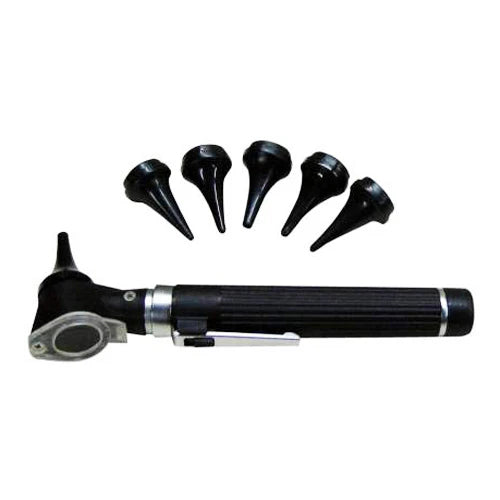
Basic Student Pocket Scope, Otoscope & Auriscope
Collection:
Professional Otoscopes & Auriscopes: ENT & Clinic Diagnostic Sets
Defining the Instrument: What is the Correct Definition for Otoscope?
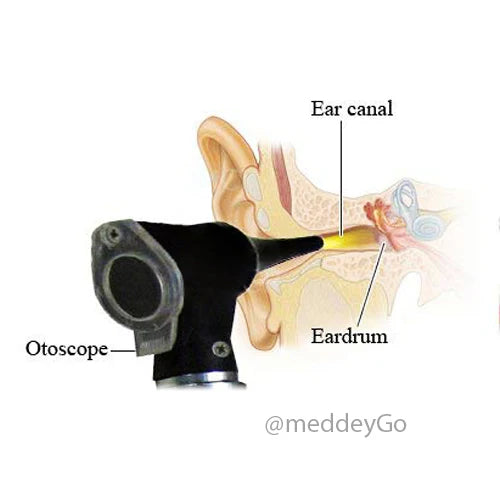
The otoscope is a specialized medical device used by healthcare professionals to visualize the ear canal and tympanic membrane. While often used interchangeably with the term auriscope, the clinical community views the otoscope as an essential gateway for diagnosing conditions of the outer and middle ear.
An otoscope instrument typically consists of three primary components: a handle (containing the power source), a head (containing the light source and magnifying lens), and a speculum (the disposable or reusable tip that enters the ear canal). Modern otoscopes for doctors now utilize Fiber Optics and LED illumination to provide a "Cool Light" spectrum, ensuring that the delicate tissues of the ear are not subjected to heat during prolonged examinations.
Pro-Tip: Auriscope vs. Otoscope
Technically, an auriscope is a broader term for any device used to inspect the ear. However, in modern medical procurement, the term otoscope is preferred as it implies the presence of a magnifying lens and an integrated light source, which are standard in today’s diagnostic sets.
What is an Otoscope Used For?
The primary answer to what is an otoscope used for is the screening and diagnosis of ear pathologies. By providing a clear, illuminated view of the external auditory canal and the eardrum, the device allows for the detection of Otitis Media (middle ear infection), Otitis Externa (swimmer's ear), and Cerumen Impaction (wax buildup).
Beyond basic inspection, otoscopes for doctors are used for:
- ◈ Foreign Body Removal: Providing the illumination needed to safely extract objects using forceps.
- ◈ Tympanostomy Tube Monitoring: Checking the placement and patency of ventilation tubes in pediatric patients.
- ◈ Nasal Examination: Many clinicians use an otoscope instrument with a larger speculum for a cursory view of the nasal turbinates.
Speculum Size Compatibility Table
(Hover over sizes to see typical patient demographic use)
| Speculum Size (Diameter) | Patient Type | Clinical Goal |
|---|---|---|
| 2.5 mm | Infant / Pediatric | Deep visualization of tiny ear canals |
| 3.0 mm | Adolescent | Standard diagnostic screening |
| 4.0 mm | Adult | Maximum light entry and wide field of view |
| 5.0 mm | Adult / Large Canal | Ideal for foreign body extraction |
The Diagnostic Spectrum: What are the Types of Otoscopes?
In a high-traffic clinic or hospital, the choice of otoscope instrument depends on the mobility of the practitioner and the complexity of the patient's needs. While all otoscopes for doctors share the same basic function, their form factors vary significantly to accommodate different medical environments.
Wall-Mounted Otoscopes
Standard in ERs and exam rooms. These are connected to a permanent power source, ensuring the otoscope is always ready for high-volume use without battery failure.
Pocket Otoscopes
The preferred auriscope for residents and physios. Lightweight and battery-powered, these fit in a lab coat pocket while maintaining high-intensity LED light.
Fiber Optic Otoscopes
Unlike direct-illumination models, these use fiber optic bundles to project light from the handle to the tip, providing an unobstructed, shadow-free view of the ear canal.
The evolution of the otoscope instrument has also led to the "Video Otoscope" or ear camera used by ENT specialists. These digital models allow for live streaming of the ear canal onto a monitor, facilitating patient education and high-resolution documentation for surgical planning.
The Digital Frontier: What is the Ear Camera Used by ENT?
Modern Otolaryngology has been transformed by the ear camera used by ENT specialists. Formally known as a digital video otoscope, this device replaces the traditional eyepiece with a high-definition CMOS or CCD sensor. This allows the otoscope instrument to capture photos and videos of the tympanic membrane in real-time.
Advanced Diagnostics: The Pneumatic Otoscope
For a definitive diagnosis of middle ear fluid, the pneumatic otoscope is an indispensable tool. This version of the otoscope for doctors includes an airtight lens and a specialized attachment for a rubber bulb.
In a healthy ear, the tympanic membrane should move visibly in response to the air pressure. If the eardrum remains stationary, it is a hallmark sign of "Otitis Media with Effusion" (fluid behind the drum). This otoscope examination is the gold standard for differentiating between a simple red eardrum and a true infection requiring antibiotics.
Clinical Mastery: The Otoscope Examination Protocol
Performing a professional otoscope examination is an art form that balances patient comfort with diagnostic precision. Whether you are using a pocket auriscope or a high-end wall unit, the methodology remains the same. A rushed examination often leads to missing subtle signs of middle ear disease, such as a retracted eardrum or early-stage cholesteatoma.
1. External Inspection & Palpation
Before introducing the otoscope instrument, inspect the pinna and periauricular area for redness or discharge. Palpate the tragus; tenderness here is a classic indicator of Otitis Externa, which may make the subsequent examination painful.
2. Straightening the Canal
To view the tympanic membrane, you must counteract the natural curve of the ear canal. For adults, pull the pinna upward and backward . For children, pull it straight back or slightly downward . This aligns the cartilaginous and bony portions of the canal.
3. The "Pencil Grip" Maneuver
Hold the otoscope like a pencil, with your pinky finger extended. Rest this finger against the patient's cheek. This "bracing" technique ensures that if the patient moves suddenly, the instrument moves with them, preventing accidental trauma to the ear canal.
Once the otoscope for doctors is inserted, identify the "Cone of Light" (light reflex). In a healthy right ear, it appears at the 5 o'clock position; in the left, at 7 o'clock. Absence of this reflex often indicates increased pressure or fluid in the middle ear.
Operational Safety: How to Use an Otoscope
Knowing how to use an otoscope safely is paramount, especially when dealing with uncooperative pediatric patients or adult individuals with thin canal skin. The most common error in auriscope use is inserting the speculum too deeply or with too much force.
Allow the child to "examine" a toy with the otoscope instrument first. This desensitizes them to the light and the presence of the device.
Always use the largest speculum that fits comfortably. A larger tip allows more light into the canal and provides a wider field of view for the otoscope.
Obstruction Management: The Cerumen Barrier
A common challenge during an otoscope examination is the presence of cerumen (ear wax). If the view of the eardrum is obscured by more than 50%, a definitive diagnosis cannot be made.
- [!] Visual Assessment: Use the otoscope for doctors to determine if the wax is soft, hard, or impacted.
- [!] Safe Extraction: If the clinician is trained, a curette can be used through the viewing port of some otoscope instrument models to clear a path.
- [!] Irrigation Post-Check: After ear syringing, always perform a follow-up auriscope check to ensure the drum is intact and all debris is removed.
The Light Spectrum: Otoscope Illumination Technologies
The quality of an otoscope examination is 90% dependent on light quality. In earlier decades, otoscopes for doctors relied on vacuum or halogen bulbs which produced a yellowish hue. Today, the shift toward LED and Fiber Optics has revolutionized how we view the ear canal, providing "true tissue color" which is vital for identifying subtle inflammation.
| Bulb Type | Color Temperature | Clinical Longevity | Best Use Case |
|---|---|---|---|
| Halogen / Xenon | Warm / Yellowish | 20–30 Hours | Standard primary care screening |
| LED (Standard) | Cool / Bright White | 10,000+ Hours | High-volume clinics / Portable use |
| Fiber Optic LED | Neutral / Natural | 20,000+ Hours | ENT Specialists / Surgical Prep |
For a physio or specialized clinic, LED technology is highly recommended. Not only does it provide a clearer view of the tympanic membrane, but it also consumes significantly less battery power, ensuring your auriscope is ready for use throughout a busy shift without constant recharging.
Expanding Scope: Otoscopes in Physiotherapy & General Clinics
Why is an otoscope instrument becoming a staple in non-ENT environments? Physiotherapists, especially those specializing in Vestibular Rehabilitation, use otoscopes for doctors to rule out ear-related causes of vertigo and balance disorders before beginning treatment.
Differential Diagnosis
Physios use the otoscope to ensure symptoms like dizziness aren't caused by a simple wax impaction or a visible infection before performing the Epley Maneuver.
Trauma Assessment
In sports clinics, an auriscope is used to check for tympanic membrane ruptures or barotrauma following head injuries or water-based accidents.
Geriatric Support
Ensuring hearing aid functionality by checking for wax blockages is a vital part of holistic rehabilitation in adult care units.
The Power Dilemma: Battery vs. Rechargeable Handles
When procuring an otoscope instrument, the power handle is often overlooked. However, for a hospital or clinic, this decision impacts long-term operational costs. Standard AA-battery handles are affordable upfront but lead to waste. Lithium-ion rechargeable handles, while expensive, provide consistent light intensity that doesn't "dim" as the charge depletes.
Important Note for Procurement:
If your facility uses a 3.5V head, it must be paired with a 3.5V handle. Mixing voltages between heads and handles can either underpower the bulb (leading to poor visualization) or blow the LED circuit entirely.
Always look for "Universal Handles" that allow you to swap an otoscope head for an ophthalmoscope head. This modularity is what makes a diagnostic set a lifetime investment for any doctor or clinic.
The Otoscope Masterclass: Clinical FAQ
The correct definition for otoscope is a specialized diagnostic medical instrument equipped with a light source and a low-power magnifying lens (usually 3x to 4x). Unlike a standard penlight, an otoscope instrument is designed to provide coaxial illumination. This means the light travels along the same axis as the clinician's line of sight, which is essential for visualizing the narrow, dark recesses of the ear canal. Without this specific optical alignment, shadows would obscure the tympanic membrane, making a definitive diagnosis impossible.
In a physio or clinic setting, an auriscope is used as a screening tool for vestibular and balance disorders. Since the inner ear is responsible for equilibrium, a therapist must rule out external or middle ear obstructions (like cerumen or fluid) before concluding that a patient's dizziness is neurological. An otoscope examination allows the therapist to ensure the "conducting" part of the ear is clear, ensuring that rehab protocols like the Epley Maneuver are appropriate and safe for the patient.
The ear camera used by ENT specialists is technically a Digital Video Otoscope. It utilizes a high-definition CMOS sensor to project the image of the ear canal onto a monitor. This improves outcomes by allowing for Digital Documentation (saving images for pre/post-op comparison), Patient Compliance (showing parents the infection in their child's ear), and Telemedicine , where an ENT can review high-resolution footage from a remote clinic or hospital to provide a specialist consult without the patient traveling.
The pneumatic otoscope is the only non-invasive way to measure "Tympanic Membrane Mobility." In children, middle ear fluid (effusion) is often present without pain or redness. A standard otoscope examination might miss this. By using the pneumatic bulb to change the air pressure in the canal, a doctor can see if the eardrum moves. If it doesn't, it confirms fluid is present, which is critical for deciding if a child needs ear tubes or a specific course of antibiotics to prevent hearing loss during speech development years.
When discussing what are the types of otoscopes, we categorize them by power source and illumination. For a multi-doctor hospital or clinic, Wall-Mounted Systems are best as they eliminate the "dead battery" syndrome. However, for sheer diagnostic quality, a Fiber Optic LED Otoscope is superior. It provides a shadow-free view and a 360-degree ring of light around the speculum, ensuring that the clinician can see the entire perimeter of the eardrum—a common site for subtle perforations.
Your Diagnostic Partner: The MeddeyGo Promise
At MeddeyGo, we understand that a doctor's diagnosis is only as good as the tools they use. We curate only the highest-tier otoscope instruments for the Indian medical community.
Verified Optical Quality
Every auriscope in our collection features scratch-resistant, anti-reflective lenses. We ensure that you get a crystal-clear view, session after session, without distortion.
After-Sales Support
Unlike generic marketplaces, MeddeyGo provides specialized support for bulb replacements, rechargeable battery packs, and compatible specula for all our otoscopes for doctors.
Hospital-Grade Durability
We stock brands built to survive the rigors of a 24/7 hospital environment, featuring impact-resistant casings and reinforced fiber-optic bundles.