Filter
1 product
Type: General Instruments
Uterine Sound SS Deluxe Quality (Pack of 2)
Collection:
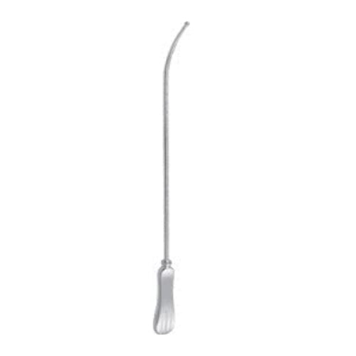
Uterine Sound Instrument: Sims & Simpson Uterine Sound – MeddeyGo
Clinical Definition: What is a Uterine Sound?
In the specialized field of gynecology and obstetrics, the uterine sound is an indispensable diagnostic and preparatory tool. Technically defined, a uterine sound is a slender, flexible or malleable probe typically constructed from surgical-grade stainless steel or silver-plated materials. The uterine sound instrument is characterized by its blunt, rounded tip and a graduated shaft that allows for the precise measurement of the uterine cavity's depth and direction.
The primary purpose of the uterine sound is to explore the cervical canal and the internal environment of the uterus. Before any intrauterine intervention can take place, the clinician must have a clear understanding of the organ's orientation—whether it is anteverted, retroverted, or mid-positioned. The uterine sound instrument provides this tactile feedback, serving as an extension of the surgeon's hands to prevent accidental trauma during more invasive procedures.
What is Uterine Sounding and Why is it Important?
Uterine sounding is the clinical procedure of inserting a sterile uterine sound through the cervical os to determine the length of the uterine cavity. This process is not merely a routine step; it is a critical safety protocol. The importance of utilizing a uterine sound includes:
- • Accurate IUD Placement: Using a uterine sound before inserting an intrauterine device ensures the provider knows the exact depth, preventing the device from being placed too low or causing perforation.
- • Procedural Safety in Surgery: For dilation and curettage (D&C), the uterine sound uses involve establishing a "safe depth" to ensure surgical instruments do not exceed the uterine fundus.
- • Pathological Screening: A uterine sound can detect physical obstructions such as submucosal fibroids, polyps, or congenital septums that might interfere with fertility or menstrual health.
Technical Architecture: Uterine Sound Parts and Names
Understanding the uterine sound parts name is essential for effective communication in the operating theater. While the instrument appears simple, each component of the uterine sound instrument is engineered for a specific function to ensure patient comfort and measurement accuracy.
| Component Name | Technical Description | Function of the Part |
|---|---|---|
| The Bulbous Tip | A smooth, rounded distal end. | Prevents the uterine sound from snagging on cervical tissue or piercing the fundus. |
| The Malleable Shaft | The long, slender middle section. | Allows the uterine sound instrument to be bent to match the patient's unique anatomy. |
| Graduated Markings | CM or Inch etchings on the shaft. | The uterine sound uses these markings to provide an objective measurement of cavity depth. |
| The Handle | A widened, often textured proximal end. | Provides the clinician with a secure grip to manipulate the uterine sound with precision. |
When discussing the parts of uterine sound, it is important to note the difference between a rigid sound and a malleable one. Malleable uterine sound instrument models are preferred in modern practice because they can be manually curved to bypass a stenotic cervix or follow the sharp angle of a retroverted uterus. This flexibility is what makes the use of uterine sound safe across a diverse patient demographic.
Differentiating Instruments: Sounds, Dilators, and Forceps
In a standard gynecological tray, the uterine sound is often placed alongside other instruments that may look similar but serve very different purposes. It is vital for surgical technicians and clinicians to distinguish the uterine sound instrument from dilators or forceps.
What is a uterine sound dilator used for? In many contexts, this refers to a multi-purpose tool or the sequential use of instruments. While the uterine sound measures depth, a dilator (like a Hegar or Pratt) is used to physically widen the cervical canal. Sometimes, a graduated dilator is used for "sounding," but a dedicated uterine sound is thinner and safer for initial exploration.
What is the use of uterine sound forceps? This is often a misnomer in clinical settings. Forceps (such as tenaculum forceps) are used to grasp and stabilize the cervix, whereas the uterine sound is used to enter the cavity. The tenaculum and the uterine sound instrument work in tandem: the forceps provide traction to straighten the cervical-uterine angle, facilitating the smooth entry of the sound.
Surgical Methodology: The Uterine Sounding Procedure
The clinical procedure of using a uterine sound is a fundamental skill in gynecology, yet it requires extreme precision and an acute sense of tactile resistance. Because the uterine sound instrument is used to probe an organ that is not directly visible, the clinician must rely on "haptic feedback" to ensure the tool remains within the safe confines of the uterine cavity.
The patient is placed in the dorsal lithotomy position. A sterile speculum is inserted to visualize the cervix. The cervix is then cleansed with an antiseptic solution. Before the uterine sound is introduced, a bimanual examination must be performed to determine if the uterus is anteverted or retroverted.
A tenaculum is typically applied to the anterior lip of the cervix (for an anteverted uterus). Gentle traction is applied to straighten the angle between the cervical canal and the uterine body. This stabilization is crucial for the use of uterine sound, as it minimizes the risk of the instrument creating a false passage.
The sterile uterine sound is gently held like a pen. It is inserted through the cervical os and advanced slowly. When the clinician feels a slight, firm resistance, the tip of the uterine sound instrument has reached the fundus.
The clinician notes the marking on the uterine sound shaft that aligns with the external cervical os. This measurement is immediately recorded. The uterine sound instrument is then slowly withdrawn, checking for any blood or discharge on the tip which could indicate underlying pathology.
Comparative Analysis: Simpson vs. Sims Uterine Sound Models
Not all uterine sound instrument designs are identical. Depending on the clinical objective—whether it is simple measurement for an IUD or preparation for a major surgery—the clinician must choose between the two primary variations: the Simpson and the Sims uterine sound.
| Specification | Simpson Uterine Sound | Sims Uterine Sound |
|---|---|---|
| Design Profile | Slightly curved distal end; rigid handle. | Straight shaft; highly malleable. |
| Primary Application | General outpatient diagnostic use. | Surgical theatre; D&C preparation. |
| Flexibility | Moderate; holds its shape well. | High; can be significantly re-shaped. |
| Measurement Markings | Standard CM or Inch graduations. | Deep-etched graduations for surgical visibility. |
The Simpson uterine sound is often the "workhorse" of the gynecology clinic. Its gentle curve is designed to naturally follow the anteverted path of the majority of patients. However, for specialized cases involving congenital malformations or severe fibroids, the Sims uterine sound is often preferred because its malleability allows the surgeon to "sculpt" the uterine sound instrument to the exact path identified during ultrasound or bimanual palpation.
Safety Protocols: Managing Discomfort and Perforation Risks
The use of uterine sound is generally considered a low-risk procedure, but it is not without potential complications. The most significant risk associated with the uterine sound instrument is uterine perforation. This occurs when the tip of the sound passes through the uterine wall into the peritoneal cavity.
To minimize this risk, the clinician must always use "gentle pressure" only. If the uterine sound does not advance with the weight of two fingers, it should be withdrawn. Perforation is more common in a recently pregnant (postpartum) uterus or in postmenopausal patients where the uterine wall is thin.
Patient discomfort is the second most common challenge. The hysterometer instrument passing through the internal os often triggers uterine cramping (prostaglandin release). To manage this, clinicians should explain the procedure clearly, use a warmed hysterometer, and sometimes apply a local anesthetic (like lidocaine spray or a paracervical block) if the patient has a high sensitivity or a history of painful cervical procedures.
Specialized Utility: Uterine Sound Uses in Fertility and Oncology
Beyond the standard IUD insertion, the hysterometer plays a pivotal role in advanced reproductive medicine and gynecological oncology. In the context of fertility treatments, specifically Embryo Transfer (ET) or Intrauterine Insemination (IUI), the hysterometer instrument is often used during a "mock transfer." This allows the reproductive endocrinologist to map the "topography" of the uterine cavity, identifying any hidden curves or obstructions that could impede the delicate placement of an embryo.
In oncology, the uterine sound is utilized to assess the extent of endometrial thickness or to locate the site of a suspected lesion before a directed biopsy. The use of hysterometerin these high-stakes environments requires a specialized "touch." Clinicians often prefer silver-plated sounds for these tasks, as they offer a higher degree of malleability than standard stainless steel, allowing the hysterometer instrument to glide over fragile, potentially neoplastic tissue without causing iatrogenic bleeding.
What is Uterine Sounding Important for in Post-Menopausal Care?
For post-menopausal patients presenting with abnormal uterine bleeding, the hysterometer is a primary diagnostic gatekeeper. Because the uterus undergoes atrophy after menopause, the cavity can become significantly smaller and the cervix more rigid. Using a hysterometer instrument allows the clinician to determine if the cavity is even accessible for a Pipelle biopsy or if a more formal surgical dilation is required under anesthesia.
Precision Ergonomics: The Anatomy of the Uterine Sound Handle
While much focus is placed on the distal tip, the handle of the hysterometerinstrument is where the clinician’s control originates. Professional-grade sounds, such as those provided by Meister Surgical, feature handles designed for maximum tactile feedback. When the hysterometer makes contact with the fundus, the vibration is transmitted through the shaft to the handle.
| Handle Feature | Technical Specification | Clinical Benefit |
|---|---|---|
| Thumb Rest / Grip | Flat or textured indentation. | Ensures the hysterometer does not rotate during insertion. |
| Weight Balance | Neutral center of gravity. | Prevents hand fatigue during complex hysterometer procedures. |
| Matte Finish | Anti-glare surgical coating. | Prevents visual interference under high-intensity OR lights when reading parts of hysterometer graduations. |
| Malleability Index | High-purity alloy composition. | Allows the hysterometer instrument to retain its custom shape without "springing back." |
The parts name for the handle area often includes the "stop" or "collar." Some specialized versions of the uterine sound feature an adjustable slide-rule collar. Once the sound reaches the fundus, the clinician slides the collar to the external os, "locking in" the measurement. This eliminates the need to remember a number and allows the clinician to show the depth to a colleague or student for verification.
Technical Differentiation: Why Sounding is Not Dilation
A common point of confusion for junior residents is the difference between a hysterometer and a cervical dilator. While both are inserted into the cervical canal, their mechanical principles are polar opposites.
The hysterometer is for information gathering. It is a diagnostic probe. Conversely, a dilator is a therapeutic or preparatory tool used to exert radial force to stretch the cervical fibers. Utilizing a dilator to measure depth is inherently dangerous because dilators are typically thicker and more rigid; they require more force to pass through the internal os, which masks the subtle "fundal tap" that a hysterometer provides.
What is a uterine sound instrument used to measure accurately?
Accuracy in hysterometer is defined by the instrument's ability to reach the fundus without displacing it. A heavy dilator may actually stretch the uterus or push it upward, resulting in an artificially long measurement. The lightweight, slender profile of a hysterometer ensures that the measurement recorded is the true anatomical depth, which is the gold standard for surgical safety.
Conclusion: The Enduring Role of the hysterometer
The uterine sound remains a cornerstone of gynecological practice, bridging the gap between external examination and internal intrauterine intervention. Throughout this masterclass, we have identified that the uterine sound instrument is far more than a simple measuring rod; it is a vital safety diagnostic that dictates the success of IUD insertions, surgical dilations, and fertility mappings. By providing an objective measurement of uterine depth and a tactile assessment of cervical patency, the uterine sound prevents the most dreaded complications in uterine surgery—perforation and false passage formation.
Modern advancements in material science have further refined the hysterometer, introducing malleable alloys and high-contrast etched graduations. These developments ensure that the hysterometer is safer and more precise than ever before. Whether a clinician is using a Simpson model for routine diagnostics or a Sims model for complex surgical preparation, the fundamental principle remains the same: the uterine sound acts as the "eyes" of the clinician inside the uterine cavity.
Why Uterine Sounding is Important in Modern Medicine
In an era of high-tech imaging, one might ask if the manual hysterometer is still necessary. The clinical answer is a definitive yes. While ultrasound can provide a visual estimate, the hysterometer provides real-time tactile confirmation of the fundal wall's integrity. It is the only tool that allows a clinician to physically confirm that the path through the cervix is clear and ready for subsequent instrumentation. This makes the hysterometer an irreplaceable component of any gynecological tray.
Maintenance & Sterilization of the Uterine Sound
To maintain the longevity and safety of your hysterometer, strictly adhere to these surgical-grade decontamination protocols:
- • Pre-Cleaning: Immediately after use, rinse the hysterometer in cold water to prevent blood from drying in the etched markings.
- • Ultrasonic Cleaning: Use an ultrasonic bath to dislodge microscopic debris from the hysterometer graduations.
- • Autoclaving: Standard steam sterilization (134°C for 3 minutes) is the gold standard for stainless steel hysterometer models.
- • Inspection: Before every use, check the hysterometer for burrs, nicks, or loss of malleability that could lead to uterine trauma.