


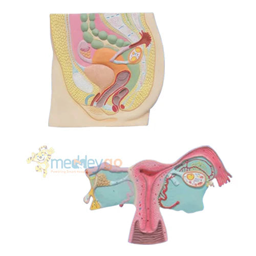


Female Reproductive System 3D Model with 56 Labels
Female Reproductive System 3D Model with 56 Labels
In stock
- Free shipping over ₹500
Couldn't load pickup availability
Share this product
Female Reproductive System 3D Model with 56 Marked Structures
The Female Reproductive System 3D Model with 56 Marked Structures is designed for medical colleges, nursing institutes, hospitals, gynecology departments and anatomy laboratories. This female reproductive system 3D model provides a clear sectional view of the reproductive organs and nearby pelvic structures. Therefore, learners can understand how the uterus, ovaries, fallopian tubes, bladder and rectum relate to one another.
The model includes two detailed teaching sections. The upper section shows the female pelvis with 20 marked anatomical positions. In addition, the lower section focuses on the uterus and its adnexa with 36 marked positions. As a result, instructors can begin with the complete pelvic arrangement and then explain smaller reproductive structures in greater detail.
Advanced HSP resin provides a firm and durable teaching surface. Moreover, the colour-coded finish helps users separate organs, vessels, ducts and nearby tissues. Its large 40 × 80 cm format improves visibility during classroom demonstrations and clinical presentations.
Key Features
- Detailed 3D Anatomy: Presents internal and external female reproductive structures in a clear sectional form.
- Two Teaching Sections: Combines a complete pelvic view with a focused uterus and adnexa section.
- 56 Marked Positions: Includes 20 marked structures in the upper section and 36 in the lower section.
- Uterus and Cervix Detail: Shows the uterine cavity, cervix and vaginal folds for reproductive anatomy study.
- Ovaries and Fallopian Tubes: Displays both ovaries, uterine tubes and fimbriae in their anatomical relationship.
- Pelvic Organ Context: Includes the bladder, urethra, rectum and surrounding structures.
- Colour-Coded Finish: Makes nearby tissues and organs easier to identify during teaching.
- Durable HSP Resin: Supports repeated handling in colleges, clinics and training laboratories.
Technical Specifications
| Specification | Details |
|---|---|
| Product Type | Female Reproductive and Pelvic Anatomy Model |
| Display Format | Three-Dimensional Sectional Anatomy Model |
| Number of Sections | 2 Detailed Teaching Sections |
| Total Marked Positions | 56 |
| Upper Section | Female Pelvis with 20 Marked Anatomical Positions |
| Lower Section | Uterus and Adnexa with 36 Marked Positions |
| Structures Shown | Uterus, Cervix, Vagina, Ovaries, Fallopian Tubes, Fimbriae, Bladder, Urethra, Labia and Rectum |
| Material | Advanced HSP Resin |
| Approx. Size | 40 × 80 cm |
| Usage | Medical Education, Anatomy Teaching and Patient Counselling |
Benefits
- First, students can study reproductive and pelvic organs together.
- Next, the two sections support both general and detailed learning.
- Also, marked positions make structure identification easier.
- In addition, colour coding improves visual understanding.
- Therefore, teachers can explain organ relationships more clearly.
- Moreover, the large format supports group demonstrations.
- Finally, durable resin supports regular institutional use.
Suitable For
- Medical colleges and universities
- Nursing and midwifery institutes
- Gynecology and obstetric departments
- Anatomy and physiology laboratories
- Hospitals, clinics and training centres
- Patient education and counselling rooms
Applications
- Female reproductive anatomy teaching
- Pelvic organ relationship study
- Uterus and cervix demonstrations
- Ovary and fallopian-tube education
- Medical and nursing practical classes
- Reproductive-health counselling
Why Choose This Female Reproductive System 3D Model?
This female reproductive system 3D model combines a full pelvic view with a focused reproductive section. For example, instructors can first show the position of the uterus between the bladder and rectum. Then, they can use the lower section to explain the ovaries, fallopian tubes and uterine cavity. Consequently, learners gain a connected understanding of female pelvic anatomy.
The 56 marked positions also support organised lessons and revision. In addition, the large design makes important structures easier to see. Therefore, the model works well for lectures, practical examinations and patient education.
Buying Guidance
Before buying, confirm that your course requires both pelvic and reproductive-system anatomy. Also, check the clarity of the marked structures, anatomical colouring and overall finish. Finally, confirm whether a numbered identification guide is included with the supplied model.
Usage Note: Use this model only for education and demonstration. It cannot diagnose reproductive or pelvic conditions or replace clinical examination, ultrasound, imaging and professional medical advice.
FAQs
1. What does this female reproductive system model show?
It shows the uterus, cervix, vagina, ovaries, fallopian tubes, bladder, urethra, labia and rectum.
2. How many anatomical positions are marked?
The model includes 56 marked positions across its two sections.
3. Does it show the ovaries and fimbriae?
Yes. Both ovaries, the fallopian tubes and their fimbriae are shown.
4. What material is used?
The model is made from durable HSP resin.
5. What is the approximate size?
The supplied size is approximately 40 × 80 cm.
6. Is it suitable for gynecology teaching?
Yes. It supports gynecology, obstetric, nursing and reproductive-health education.
7. Can it be used for patient education?
Yes. Healthcare professionals can use it to explain female reproductive anatomy visually.
